Published: August 2018
Atrial fibrillation (AF) is a common heart rhythm disorder caused by degeneration of the electrical impulses in the upper cardiac chambers (atria) resulting in a change from an organized heart rhythm to a rapid, chaotic rhythm. The resulting arrhythmia is often rapid and irregular with no discernible pattern (known as irregularly irregular). The disrupted rhythm occurs because of the unpredictable conduction of disordered impulses across the electrical bridge, called the atrioventricular (AV) node, to the lower cardiac chambers (ventricles). The arrhythmia also results in ineffectual atrial contractions affecting cardiac output and vulnerability to blood clot (thrombus) formation that can result in stroke events.
According to the 2014 American Heart Association (AHA)/American College of Cardiology (ACC)/Heart Rhythm Society (HRS) clinical practice guidelines, AF can be classified based on the duration of episodes. Paroxysmal AF refers to AF that begins suddenly and ends spontaneously within 7 days of onset.1 Persistent AF refers AF that occurs for longer than 7 days and ends spontaneously or with treatment. Long-standing persistent AF refers to patients who have uninterrupted AF for more than a year. Permanent AF refers to AF that persists despite treatment to restore normal sinus rhythm or that is not treated.
These AF classifications are not mutually exclusive and it is common for patients with one type of AF to exhibit overlapping features of another type. These classifications are relevant clinically with respect to outcomes and prognosis with rhythm-controlling treatment strategies. Atrial fibrillation is commonly associated with other supraventricular arrhythmias, namely atrial flutter and focal atrial tachycardia.
Atrial fibrillation is the most common sustained cardiac tachyarrhythmia encountered by clinicians worldwide. An estimated 2.7 to 6.1 million people in the United States have Atrial fibrillation with projections to reach nearly 12.1 million in 2030.1,2
The prevalence of A fib increases with age, afflicting about 10% of the population by 80 years of age.3 The growing prevalence of AF may be influenced by extended survival outcomes for patients with congestive heart failure (CHF), valvular heart disease, and coronary artery disease as AF is common among patients with other forms of structural heart disease.
Atrial fibrillation may be acutely associated with physiologic stressors such as surgical procedures, pulmonary embolism, chronic lung diseases, hyperthyroidism, and alcohol ingestion. Disease states commonly associated with AF include hypertension, valvular heart disease, CHF, coronary artery disease, Wolff-Parkinson-White syndrome, pericarditis, obstructive sleep apnea, and cardiomyopathy. Considerable research has been devoted to the mechanisms and pathogenesis of AF. Genetic studies have identified specific associations, particularly in the cases of familial AF 4 Achieving a complete understanding of AF is limited by the complexity of this disorder and the heterogeneous patient population it affects.
The pathogenesis of AF can be broadly divided into the categories of triggers, substrate, and sustaining mechanisms. Since the late 1990s, it has been recognized that the initiation of AF can occur because of premature atrial contractions triggered by beats that arise from the pulmonary veins (PVs), usually from muscular tissue sleeves near the junction with the left atrium.5 These triggers may also fire repetitively and contribute to the maintenance of AF, essentially becoming drivers of AF. Focal triggers outside the PV including posterior left atrial, ligament of Marshall, coronary sinus, venae cavae, septum, and left atrial appendage contribute to the disease process. Focal triggers, especially the PVs, are felt to be very important early in the disease process and, in particular, among patients with paroxysmal AF. Over time, myocardial fibrosis develops within the atrial tissue in association with AF to support its maintenance by shortening affected tissue refractory periods. Myocardial fibrosis of the atrium seems to be the common feature of the progression of AF disease state. This has led to the adage that AF begets AF. Once AF is initiated by focal triggers, several theories have been postulated to explain the maintenance of AF. They include the multiple wavelet model, AF rotors and the role of the autonomic nervous system. The multiple wavelet model has suggested that AF is sustained by multiple simultaneous wavelets meandering throughout the atria. Atrial tissue with abnormal electrical propagation recorded by mapping catheters has been referred to as complex fractionated electrograms. Expression of specific connecting protein channels at the cellular level are also felt to be important contributors to the disease substrate and sustaining mechanisms. Contemporary understanding of the AF substrate and sustaining mechanisms now also includes the role of the autonomic nervous system and, more recently, the discovery and evaluation of the concept of AF rotors.6,7
Cardiac ganglionic plexuses clustered posteriorly and superiorly to the left atrium are known to play an important role in the initiation and maintenance of AF. Both parasympathetic and sympathetic limbs can provoke atrial arrhythmias. Evidence supportive of this concept includes therapeutic benefit derived from destruction of cardiac ganglionic plexuses and also noncardiac plexuses including the stellate ganglion and perinephric ganglia associated with the renal arteries.8 In addition, completely vagally denervated hearts as in heart transplantation are known to have a very low incidence of AF.9
Atrial fibrillation rotors represent an emerging concept as a sustaining mechanism for AF involving spiral waves detected by spectral analysis of dominant frequencies recorded by intracardiac mapping catheters. Such spiral waves can be conceptualized as wavelets of consistent electrical activation around a central localized source that could be either structural (ie, scar-related) or purely functional (ie, conduction heterogeneity involving certain cellular sodium and potassium channels). The focal impulse and rotor modulation computational mapping system is used to identify AF rotors.10
Atrial fibrillation may have hemodynamic consequences. It can decrease cardiac output due to ineffectual atrial systole and increase pulmonary venous pressure resulting in heart failure. Deleterious hemodynamic effects also include nonphysiologic tachycardia, increased valvular regurgitation, and irregularity in ventricular systole.
AF is associated with morbidity and even mortality. AF can produce bothersome symptoms that affect quality of life, but patients with AF also have a substantial risk of thromboembolic stroke, AF is associated with a fivefold increased risk of stroke, threefold risk of heart failure. and twofold risk of dementia and mortality.1 Some data demonstrate an association of AF with reduced overall survival.11
The clinical manifestations of AF are variable, although fatigue is the most common symptom. Often, the symptoms are attributable to the rapid ventricular response. However, even when the ventricular response is controlled, symptoms can occur from loss of AV synchrony or atrial systole This is particularly important for patients with left ventricular dysfunction (CHF) and impaired diastolic filling (mitral stenosis , hypertrophic and restrictive cardiomyopathy) .That said, some patients with AF are genuinely asymptomatic, even at rapid heart rates for unclear reasons. More often, however, patients report nonspecific symptoms such as fatigue, dyspnea, dizziness, and diaphoresis. Palpitations are a common feature. Occasionally, patients present with extreme manifestations of hemodynamic compromise, such as chest pain, pulmonary edema, or syncope. Atrial fibrillation is present in 10% to 40% of patients with a new thromboembolic stroke.12
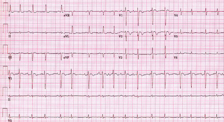
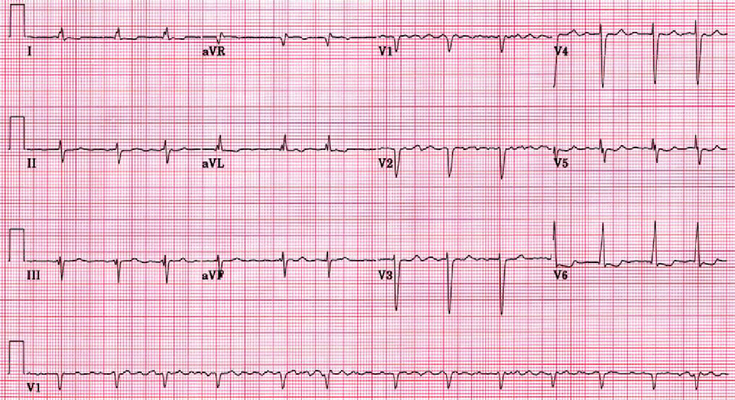
The clinician must realize that an irregular pulse detected by physical examination or an irregular ventricular rhythm seen on the electrocardiogram (ECG) is not always AF. It is necessary to consider and exclude other types of irregular rhythm disturbances, including atrial or ventricular ectopy, atrial tachycardia, or atrial flutter (Figure 1) with variable AV conduction, multifocal atrial tachycardia (Figure 2), and wandering atrial pacemaker. Conversely, a regular pulse or rhythm does not exclude AF. For example, AF can manifest with a regular ventricular response in the presence of AV block or with a ventricular paced rhythm.
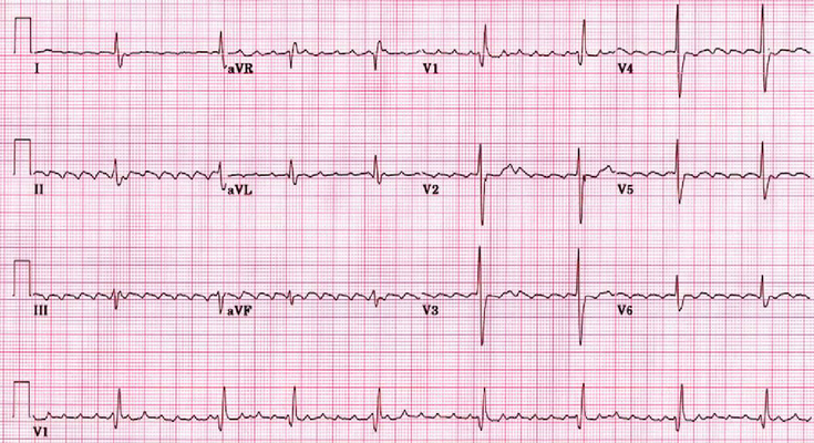
A 12-lead ECG is best to establish the diagnosis of AF. Electrocardiographic findings in AF include the absence of P waves and the presence of low-amplitude, high-frequency atrial fibrillary waves (F waves). The atrial rate varies in the range of 300 to 700 beats per minute. In the absence of drug therapy, a patient with normal AV conduction has an irregularly irregular ventricular rhythm and often has a ventricular rate in the range of 120 to 180 beats per minute.13 The baseline on the ECG strip often is undulating and occasionally has coarse irregular activity (Figure 3). This activity may resemble atrial flutter, but it is not as uniform wave to wave as atrial flutter.
Most patients presenting with AF are not critically ill. However, in some cases, the presence of AF may cause life-threatening hemodynamic compromise. It should be emphasized that for any unstable patient presenting with AF—for example, a patient with chest pain, pulmonary edema, or hypotension—the recommended therapy is rapid electrical cardioversion, according to the Advance Cardiovascular Life Support guidelines.14
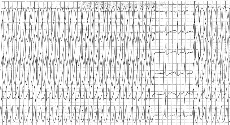
Atrial fibrillation has particular importance in the setting of the Wolff-Parkinson-White syndrome. Patients with Wolff-Parkinson-White syndrome may be vulnerable to ventricular fibrillation and sudden death because of the development of AF, which can result in extremely rapid conduction over the accessory pathway (Figure 4). Prompt electrical cardioversion is of utmost importance for these patients. Treatment with AV node-blocking medications such as verapamil or digoxin can facilitate rapid conduction over the accessory pathway and result in ventricular fibrillation. When intravenous pharmacologic therapy is required, the drug of choice is procainamide or amiodarone.
There are 3 goals in the management of AF: control of the ventricular rate, minimization of thromboembolism risk (particularly stroke), and restoration and maintenance of sinus rhythm. The first 2 goals are essential for most patients, but the third goal may not be necessary in all patients. The AHA/ACC/HRS guidelines provide a more detailed review of the management of patients with AF treated purely with a rate-controlling approach and those patients treated to restore and maintain normal sinus rhythm (ie, a rhythm controlling strategy).1
Rate control in patients with AF is essential to reduce symptoms and improve quality of life. The optimal heart rate goal has not been fully defined and may be patient specific. In the RACE II clinical trial, patients were randomly assigned to strict (less than 80 bpm) vs lenient (less than 110 bpm) rate control strategies.15 Lenient rate control was not inferior to strict rate control in terms of cardiovascular morbidity and mortality. Based on this study, the European Society of Cardiology guidelines incorporated the lenient rate control strategy as the first-line approach to asymptomatic patients with preserved cardiac function.16 However, the guidelines from AHA/ACC/HRS society favor a more stringent rate control strategy (class II A recommendation).
Guideline statements only address goals in patients with preserved cardiac function. The optimal rate in patients with heart failure has not been fully defined. For example, some studies show that in patients with heart failure, slow ventricular rates are associated with higher mortality and higher ventricular rates may be needed to improve exercise tolerance.17,18 However, patients with heart failure can easily become decompensated when ventricular rates are uncontrolled. Hence, most clinicians use a patient-specific window of optimal rate control that avoids the consequences of both extreme bradycardia and tachycardia.19
Ventricular slowing is accomplished with medications affecting the AV node (Table 1). The most commonly used drug classes are beta blockers and calcium channel blockers. Most patients with persistent atrial fibrillation receive daily suppressive therapy. However, a pill-in-the-pocket, rate-control strategy has been proposed in patients with a low burden of self-terminating AF, though no studies have investigated this strategy.19
If these medications are ineffective or if excessive bradycardia occurs, other measures may need to be considered. One option suitable for some patients is catheter ablation of the AV node and pacemaker implantation (ablate and pace). Meta-analysis of the ablate-and-pace approach has demonstrated improvements in a number of clinical parameters, including symptoms, quality of life, exercise function, cardiac performance, and longevity in patients with CHF receiving a biventricular pacemaker.20
However, this approach usually results in pacemaker dependence and carries the associated risks and complications of indwelling pacemaker leads. Pacemaker implantation without AV nodal ablation should be considered if the problem is simply excessive bradycardia that prohibits the effectiveness of rate-controlling medication, and the rapid ventricular rates are well controlled by medication. For patients with abnormal LV systolic function, a biventricular pacemaker (ie, cardiac resynchronization therapy) should be considered in conjunction with AV nodal ablation based on the results of the BLOCK HF trial.21
| Beta Blockers • Advantages: Intravenous administration produces a rapid onset of effect; heart rate control at rest and with activity; oral forms available. • Disadvantages: May worsen heart failure in decompensated patient. May exacerbate reactive airway diseases; cause fatigue, depression, and impotence. Abrupt withdrawal may cause rebound tachycardia, hypertension and myocardial ischemia. |
|||
| Drug | Dose | Onset of Action | Duration of effect |
|---|---|---|---|
| Propranolol | Intravenous | 1 mg bolus, repeat every 5 minutes as needed to achieve goal | 5 minutes | 1 to 4 hours |
| Oral immediate release | 10 to 30 mg every 6 or 8 hours daily | 1 to 2 hours | 6 to 24 hours depending on form |
| Oral extended release | 80 to 160 mg daily | 1 to 2 hours | 6 to 24 hours depending on form |
| Metoprolol | |||
| Intravenous | 2.5 to 5 mg every 2 to 3 minutes as need to achieve goal | 5 minutes | 1 to 3 hours |
| Oral immediate release | 12.5 to 100 mg every 6 to 8 hours | 1 to 2 hours | 3 to 24 hours depending on form |
| Oral extended release | 50 to 400 mg once daily | 1 to 2 hours | 3 to 24 hours depending on form |
| Esmolol | |||
| Intravenous | 500 µg/kg over 1 minute, then maintenance dose of 25 to 300 µg/kg/min; titrate by 25 to 50 µg/kg/min every 5 to 10 minutes to achieve goal | 2 to 10 minutes | 9 minute half-life |
| Nadolol | Oral (avoid in patients with renal dysfunction) | 10 to 240 mg daily for rate control | 1 to 2 hours | 17 to 24 hours |
| Calcium Channel Blockers • Advantages: Intravenous administration produces a rapid onset of effect; heart rate control at rest and with activity; oral forms available. • Disadvantages: May worsen heart failure in decompensated patient; may cause fatigue, edema and constipation. |
|||
| Drug | Dose | Onset of Action | Duration of effect |
|---|---|---|---|
| Diltiazem | |||
| Intravenous | 0.25 mg/kg over 2 minutes, then 5 to 15 mg/hour; may repeat bolus of 0.35 mg/kg if goal heartrate not achieved | 5 minutes | 1 to 10 hours |
| Oral immediate release | 30 to 120 mg every 6 to 8 hours (typical maximum of 360 mg daily) | 1 hour | 5 to 24 depending on form |
| Oral extended release | 120 to 360 mg once daily | 1 hour | 5 to 24 depending on form |
| Verapamil | |||
| Intravenous | Initial bolus: 0.075 to 0.15 mg/kg (usual dose: 5 to 10 mg) over at least 2 minutes; if no response, may give an additional 10 mg bolus after 15 to 30 minutes | 3 to 5 minutes | 0.5 to 6 hours |
| Oral immediate release | 80 to 120 mg every 8 to 12 hours to a maximum of 480 mg daily | 1 to 2 hours | 6 to 8 hours |
| Oral extended release | 180 to 480 mg once daily | 1 to 2 hours | 6 to 8 hours |
| Other • Advantages: Can be used in patients with heart failure. Useful in combination with other atrioventricular nodal agents. • Disadvantages: Slow onset of action; poor control of heart rate with activity; narrow therapeutic margin; long duration of effect. Not recommended as monotherapy under practice guidelines. |
|||
| Drug | Dose | Onset of Action | Duration of effect |
|---|---|---|---|
| Digoxin | |||
| Intravenous | Loading dose of up to 1 mg in first 24 hours, with bolus of 0.25 to 0.5 mg; then remainder in divided doses 6 to 8 hours | 5 to 60 minutes | 3 to 4 days |
| Oral | 0.125 to 0.25 mg daily) | 1 to 2 hours | |
In patients with an implantable cardioverter defibrillator, great care is needed in both the device programming and pharmacologic rate control in order to avoid the risk of inappropriate shocks associated with rapid ventricular rates. In patients with a cardiac resynchronization therapy device, the goal is to achieve 100 % biventricular pacing.22 If the presence of atrial fibrillation prevents achievement of this goal, further consideration to rhythm controlling strategies or AV nodal ablation is recommended.
Atrial fibrillation carries a considerable risk for thromboembolism and stroke. The Framingham study has shown that during a follow up period of 30 years, patients with nonvalvular AF had a more than fivefold risk of stroke and the risk of stroke attributed to stroke increased with age.12 Ischemic stroke can be the first manifestation of occult AF, which is also referred to as subclinical AF. Ischemic strokes related to AF tend to be associated with greater morbidity and mortality than from other causes of stroke.23 As the CRYSTAL AF and EMBRACE trials have demonstrated, AF is frequently detected in patients during post stroke cardiac rhythm monitoring in patients with cryptogenic stroke.24, 25 Some data suggests that such asymptomatic AF can carry a worse prognosis ostensibly related to the stroke risk incurred without the presence of symptoms to provide a warning.26
The annual risk of stroke may be even higher in patients with AF who have 1 or more of the following risk factors: older than 65 years, female, diabetes mellitus, hypertension, heart failure, coronary artery disease, previous stroke, or transient ischemic attack. Individual stroke risk stratification can now be calculated for patients on the basis of the presence or absence of such risk factors (ie, CHADS2 and CHADS2 VA2S2C risk scores).27 Several strategies including antithrombotic therapy with vitamin K antagonists (VKA), direct oral anticoagulation (DOAC), and antiplatelet therapy with aspirin, dipyridamole, and clopidogrel. DOAC drugs include Factor Xa inhibitors (eg, apixaban, rivaroxaban, edoxban) and direct thrombin inhibitors (eg, dabigatran).
In meta-analysis, VKAs reduced stroke or systemic thromboembolism by 64% and all-cause mortality by 26% compared with placebo.23,28,29 DOACs offers an additional risk reduction of 19% for stroke or systemic thromboembolism primarily driven by reduction in hemorrhagic stroke and 10% for mortality relative to warfarin.29 The combination of warfarin with aspirin increases the bleeding risk. Warfarin is superior to aspirin and also the combination of aspirin and clopidogrel in stroke prevention.29 Aspirin and clopidogrel is superior to aspirin alone in stroke protection among patients that are warfarin ineligible, but is associated with greater bleeding risk.30
Practice guidelines include recommendations regarding the form of antithrombotic therapy for patients with AF.1 The AHA/ACC/HRS guidelines recommend the CHA2DS2-VASc score to identify patients with AF at low, moderate, or high risk for thromboembolism. A score of 0 is considered low risk and does not require not antithrombotic therapy. A score of 2 or greater is considered high risk and antithrombotic therapy with VKAs or NOACs should be considered. A score of 1 is considered moderate risk for which antithrombotic therapy or aspirin may be considered. The goal of warfarin therapy for preventing stroke and thromboembolism from AF generally is an international normalized ratio between 2.0 and 3.0. The DOAC classes of medications do not require monitoring. Safety and efficacy have been evaluated in administrative datasets in addition to clinical trials leading to U.S. Food and Drug Administration (FDA) approval. Each DOAC drug has unique properties with respect to half-life, renal clearance, and availability of pharmacologic reversal agents.
For patients who have been in AF for more than 48 hours and are not adequately anticoagulated, electrical or pharmacologic cardioversion should be delayed until appropriate measures are taken to reduce the thromboembolic risk. There are two approaches to reduce thromboembolic risk in these patients. The conventional approach is to administer oral anticoagulation for at least 3 weeks before electrical or pharmacologic cardioversion. The second approach is transesophageal echocardiography (TEE)-guided electrical cardioversion method when cardioversion cannot be postponed or an expedited approach is preferred. In such cases, once a therapeutic level of anticoagulation has been achieved with an oral agent, intravenous heparin, or subcutaneous enoxaparin, a TEE may be performed to exclude the presence of an intracardiac thrombus. If no thrombus is seen, cardioversion may be performed. TEE can detect the presence of a thrombus in the left atrium, particularly in the left atrial appendage, which is poorly visualized on transthoracic echocardiography. The TEE-guided approach has been validated in several small multicenter trials as well as in a large, randomized, multicenter trial known as the Assessment of Cardioversion Using Transesophageal Echocardiography (ACUTE) trial.31
Oral anticoagulation should be continued after cardioversion until sinus rhythm has been maintained for at least 4 weeks to allow the atrial transport mechanism to recover. If the cardioversion was performed using the TEE-guided approach with intravenous heparin as the method of anticoagulation, it is advisable to continue intravenous heparin until therapeutic oral anticoagulation is achieved. The decision to initiate and continue anticoagulation for AF shorter than a duration of 48 hours should be based on the presence of other risk factors for thromboembolism. Subgroup analyses from RELY, ROCKET AF, and ARISTOTLE support the use of DOACs at cardioversion in addition to one nonproprietary study.32
On account of the left atrial appendage accounting for the source of most thrombi in patients with nonvalvular AF, several interventions gave been designed for left atrial appendage closure and thus stroke reduction. The Watchman device, a closure device deployed percutaneously that blocks the left atrial appendage has been approved by the FDA for stroke prevention in patients with non valvular AF who are at an increased risk of stroke and who have no contra indication to short term anti coagulation.33,34
The Lariat device is another percutaneous system for left atrial appendage exclusion; however, its labelling is not specific for stroke prevention.35
The restoration and maintenance of sinus rhythm can be beneficial for patients with bothersome symptoms. However, management of patients with asymptomatic or minimally symptomatic AF has been controversial for many years. Selecting appropriate patients for a rhythm-controlling strategy are well articulated by clinical practice guidelines.1 In addition to improving symptoms, the potential benefits of restoring and maintaining sinus rhythm include avoidance of the development of atrial cardiomyopathy from ongoing AF, improvement in heart failure, and improved overall quality of life. A rhythm-control strategy often requires the use of antiarrhythmic drugs that may have significant and even life-threatening side effects, and procedures that carry uncommon, but potentially life-threatening or disabling complications. Some nonrandomized trials have reported an increase in mortality among patients who were on long-term antiarrhythmic therapy for AF, presumably from the proarrhythmic effects of the drugs.36 In addition, several randomized studies have compared the treatment strategies of ventricular rate control or rhythm control with restoration and maintenance of sinus rhythm, albeit in older patients (mean age, 65 to 70 years) with minimal or no symptoms during AF.37,40
The Atrial Fibrillation Follow-up Investigation of Rhythm Management (AFFIRM) trial, was a large, multicenter randomized study that compared rate control with rhythm control in patients with AF.37 Both treatment strategies used appropriate anticoagulation strategies according to established guidelines. This study demonstrated that a rhythm-control strategy is no better than a ventricular rate control strategy with regard to quality of life, incidence of stroke, or mortality at a follow-up of approximately 5 years. A meta-analysis of 5 randomized controlled trials comparing rate-control with rhythm-control strategies included more than 5,000 patients and demonstrated that a rate-control strategy is not inferior to a rhythm-control strategy.41 Studies comparing catheter ablation with antiarrhythmic drug therapy as an initial approach to AF control have yielded some promising results.42,43 Ongoing, large, multicenter randomized trials like the Catheter Ablation Versus Anti-arrhythmic Drug Therapy for Atrial Fibrillation (CABANA) Trial (https://clinicaltrials.gov/ct2/show/NCT00911508) and Early Treatment of Atrial Fibrillation for Stroke Prevention Trial (EAST) (https://clinicaltrials.gov/ct2/show/NCT01288352) should provide more insight regarding catheter ablation vs antiarrhythmic drug therapy. Acutely, restoration of sinus rhythm may be achieved with either pharmacologic or electrical cardioversion. It is important to remember that electrical and pharmacologic cardioversion are no different with regard to the risk of thromboembolic stroke. Therefore, the requirements for anticoagulation apply equally to either treatment strategy and are largely dictated by the patient-specific thromboembolic risk profile discussed previously.
Electrical cardioversion is more effective than pharmacologic cardioversion. The success rate direct-current electrical cardioversion with a biphasic shock is approximately 95%.44 Electrical cardioversion should be administered with the patient under deep sedation, with cardiac and hemodynamic monitoring, and in the presence of personnel skilled in airway management. The administration of an antiarrhythmic drug may promote more successful direct current cardioversion and subsequent maintenance of sinus rhythm. Similarly, it is reasonable to add an antiarrhythmic drug for any patient who develops an early AF recurrence after direct-current electrical cardioversion and to consider a repeat attempt after the drug has been initiated and reaches steady-state blood levels.
Rates of successful immediate cardioversion by pharmacologic means have ranged from 40% to 90%, with success more likely to come for patients with AF of shorter duration.45 Unlike electrical cardioversion, pharmacologic cardioversion does not require sedation. Contemporary use of pharmacologic cardioversion in the US occurs in nonelective scenarios in the emergency department or intensive care unit, and also in stable outpatients treated with a unique type of rhythm control strategy referred to as a pill-in-the-pocket approach. Elective pharmacologic cardioversion is uncommon in the U.S given the superiority of a planned electrical cardioversion under sedation with appropriate airway management personnel on hand.
The intravenous agents approved in the US for immediate pharmacologic cardioversion of AF are procainamide, amiodarone, and ibutilide(Table 2). Amiodarone is the most commonly used drug in emergency department and intensive care unit settings. Pharmacologic conversion of AF can be achieved with oral drugs. The pill-in-the-pocket approach is sometimes used with class Ic drugs like flecainide or propafenone and may be useful for select outpatients in order to quickly abort AF episodes shortly after onset. This approach has the potential to reduce emergency department visits and hospitalizations, but must be carefully initiated and supervised. It is recommended that the first such application of this strategy is done in a monitored environment, such as an emergency department, in order to establish patient-specific safety.
| Drug | Dosing | Notes |
|---|---|---|
| Procainamide | ||
| Intravenous | 20 to 50 mg/minute or 100 mg every 5 minutes (until arrhythmia controlled, hypotension occurs, QRS complex widens by 50% or total of 17 mg/kg is given); maintenance 1 to 4 mg/minute | • Can achieve therapeutic levels quickly. • Half-life elimination: 2 to 5 hours (procainamide); 6 to 8 hours (procainamide metabolite N-acetyl procainamide [NAPA]) • Blood levels of both procainamide and NAPA need to be followed to prevent toxicity, especially in the setting of renal or hepatic insufficiency, or both • Rapid administration may cause hypotension • Up to 10% of patients with congestive heart failure may experience worsened heart failure • Procainamide is rarely used as there is no longer an oral product |
| Amiodarone | ||
| Intravenous | 150 to 300 mg given over 10 to 120 minutes, depending on tolerance of blood pressure; maintenance infusion at 0.5 to 1 mg/minute | • Can be used in patients with severe left ventricular dysfunction • Half-life elimination: 25 to 120 days • Drug interactions are common with amiodarone; careful profile review is essential • Long-term use associated with many side effectsIncidence of torsades de pointes higher than with procainamide or amiodarone |
| Oral | 600 to 800 mg daily in divided doses until 10 g total; maintenance dose 200 mg daily | |
| Ibutilide | ||
| Intravenous | 1 mg bolus; can repeat after 10 minutes if no effect; use lower doses in patients weighing < 60 kg | • Avoid use in patients with baseline prolongation of QT interval • Few extra cardiac side effects • Incidence of torsades de pointes higher than with procainamide or amiodarone • Patients must be observed for a minimum of 4 hours post dose or until QT(c) returns to baseline |
Includes information from January et al.1
Many oral agents are available for long-term maintenance of sinus rhythm in patients with AF (Table 3). Class Ia antiarrhythmic drugs (quinidine, procainamide, and disopyramide) have become less commonly prescribed than in the past because of their side effect profiles. The class Ic agents, namely flecainide and propafenone, have more favorable side effect profiles and are more commonly utilized. However, the use of these medications does have some degree of risk. The Cardiac Arrhythmia Suppression Trial (CAST) has shown that flecainide and encainide are associated with an increase in mortality when used for the suppression of ventricular arrhythmias in patients who have had a myocardial infarction with ventricular dysfunction.46As a result, there is much concern about the use of the class Ic antiarrhythmics in patients who have any type of underlying coronary artery or structural heart disease. Flecainide and propafenone are usually well tolerated and are appropriate first-line options for the treatment of AF in patients without structural heart disease, left ventricular hypertrophy, or marked pre-existing conduction disease (ie, complete left bundle branch block). Drugs such as sodium channel blockers are expected to widen the QRS duration thereby increasing vulnerability to heart block among patients with very significant pre-existing His-Purkinje system dysfunction.
| Drug | Dose | Notes |
|---|---|---|
| Quinidine | ||
| Sulfate immediate release | 200 mg every 6 hours, increase with caution | • Less negative inotropic effect • Narrow toxic-to-therapeutic ratio; interacts with many drugs, including digoxin, warfarin, verapamil; high incidence of side effects (particularly GI intolerance, neurologic side effects, thrombocytopenia) • Rate controlling medication are required prior to initiating quinidine secondary to its vagolytic properties • Diarrhea is most common and significant side effect |
| Sulfate extended release | 300 mg every 8 to 12 hours | |
| Gluconate | 324 to 648 mg every 8 hours | |
| Flecainide | 50 to 200 mg once every 12 hours | • Generally, well tolerated • Significant incidence of central nervous system, visual side effects • Avoid use in patients with coronary or structural heart disease • One of the most negative inotropic antiarrhythmics • Shown to increase mortality when used to treat and suppress ventricular arrhythmias in patients after MI • Typically need to combine with an AV nodal blocking agent |
| Propafenone | ||
| Immediate release | 150 to 300 mg every 8 hours | • Generally, well tolerated • Twice-daily extended release form results in more stable blood levels than immediate-release form • Three times daily dosing • Avoid use in patients with coronary or structural heart disease and patients with broncospastic lung disease as has significant beta blocker properties |
| Extended release | 225, 325, or 425 mg every 12 hours | |
| Disopyramide | ||
| Immediate release | 100 to 200 mg every 6 hours | • Reduce dose by one-third for adults weighing less than 50 kg • May be useful in hypertrophic cardiomyopathy because of its negative inotropic side effects • Anticholinergic effects (eg, cardiomyopathy, constipation, urinary retention) • Adjust dose in patients with renal dysfunction • Most negative inotropic of the antiarrhythmic drugs • Avoid or use with extreme caution in patients on dialysis |
| Extended release | 200 to 400 mg every 12 hours | |
| Amiodarone | 100 to 200 mg once daily | • Can be used in patients with coronary or structural heart disease • Half-life elimination (≤120 days) • Many side effects with long-term use • Baseline chest x-ray, LFTs, TFTs, should be performed • Approved only for life-threatening ventricular arrhythmias (but still often used for atrial or supraventricular arrhythmias) |
| Sotalol | 40 to 160 mg once every 12 hours | • Can be used in patients with coronary structural heart disease • Beta- blocking properties allow single-agent therapy for almost all arrhythmias • Causes QTc prolongation • Use limited by side effects related to beta-blocking properties (eg, exacerbation of reactive airway disease, depression, negative inotropy) • Renally cleared; avoid use in patients with moderate to severe renal dysfunction • Inpatient telemetry recommended for initiation of therapy |
| Dofetilide | 125 to 500 mcg every 12 hours | • Generally well tolerated; few extra cardiac effects; can be used in patients with coronary or structural heart disease • Requires monitored initiation in hospital; inpatient telemetry mandated for initiation of therapy • Causes QTc prolongation; electrocardiogram must be checked within 2 to 3 hours of administration for evidence of QTc prolongation > 15% above baseline or > 500 msec (550 msec for patients with intraventricular conduction delay) • Many drug interactions |
Based on data from January et al.1
AF, atrial fibrillation; AV, atrioventricular; ECG, electrocardiogram; GI, gastrointestinal; LFT, liver function test; MI, myocardial infarction; SR, sinus rhythm; TFT, thyroid function test
Sotalol is a class III antiarrhythmic that has beta-blocking properties and is generally well tolerated. Patients may have difficulty tolerating the beta blocker side effects, such as fatigue, and there is a potential risk of excessive bradycardia. As with other class III antiarrhythmic agents, sotalol causes QT prolongation and may result in ventricular proarrhythmia, such as torsades de pointes. Careful monitoring of renal function, electrolytes, and QT interval is recommended. In addition, initiation in hospital while on telemetry monitoring should be considered although it is not required by the FDA. Lastly, drug interactions should be carefully avoided, particularly those resulting in QT prolongation.
Dofetilide, a class III antiarrhythmic, has good efficacy rates and is one of the best tolerated antiarrhythmic drugs in terms of its side effects profile. Importantly, dofetilide has also been shown to be safe for patients with cardiomyopathy, CHF, and ischemic heart disease. Therefore, dofetilide may be considered as an alternative treatment option to amiodarone. Like sotalol, dofetilide drug causes QT prolongation that may result in proarrhythmia and rarely death if excessive and is restricted to patients without advanced renal disease. Its use has been restricted by the FDA to certified prescribers and requires monitored initiation in a hospital setting followed by structured outpatient follow-up. Unlike sotalol, however, dofetilide does not cause excessive bradycardia and thus can be administered to patients without concern for exacerbating preexisting bradycardia. Dofetilide has many potentially lethal drug-to-drug interactions, including with many commonly prescribed antibiotics and antihypertensive drugs. Despite these limitations, many patients experience improved AF control with dofeilide with fewer daily side effects compared with other antiarrhythmic drugs.
Amiodarone is generally reserved for patients with AF for whom other antiarrhythmic drugs have been contraindicated, ineffective, or poorly tolerated. This is primarily because amiodarone has potential time- and dose-dependent organ toxicities that can affect the liver, thyroid, lungs, and eyes. It is recommended that baseline tests be performed at initiation of the drug, including an ophthalmologic examination, pulmonary spirometry and diffusion capacity tests, and blood tests to assess liver and thyroid function. The blood tests are often repeated at regular intervals, approximately every 6 to 12 months, and the ophthalmologic examination should be performed yearly. Cumulative toxicity with amiodarone occurs at a rate of approximately 1% to 2% per year.
Dronedarone is an antiarrhythmic drug designed to function similarly to amiodarone but without the molecular iodine interface associated with some of the previously described amiodarone toxicities. Early enthusiasm for this drug, based on results from the initial studies, was later tempered by safety concerns and limitations. The biggest safety concern with this drug involves use in patients with CHF. It is contraindicated for patients with advanced (New York Heart Association functional class IV failure) and was found to increase cardiovascular death rates when given to patients with permanent AF in the Permanent Atrial Fibrillation Outcome Study Using Dronedarone on Top of Standard Therapy (PALLAS) study.47 This drug also has gastrointestinal side effects that can be partially mitigated by taking the drug with food. It also has some important drug-to-drug interactions, including with the anticoagulant drug dabigatran. That said, it is a reasonable treatment option for patients without structural heart disease or advanced liver disease and does not require hospital-based initiation like dofetilide. It has also been one of the most heavily studied antiarrhythmic drugs on the market.
Implantable cardiac devices are also used in the treatment of patients with AF. There is a substantial incidence of sinus node and AV node dysfunction in the AF population requiring cardiac pacing. Pacemakers have several purposes, including bradycardia pacing support, ventricular response regularization, and AF suppression or termination. Clinical practice guidelines detail the recommended uses of implantable pacemakers and anti-tachycardia devices.48 Pacemakers may be implanted simply for pacing support in patients with post-AF conversion pauses or symptomatic bradycardia while in AF. Sinus node dysfunction in association with AF is often referred to as tachycardia-bradycardia syndrome. Sinus node dysfunction can be exacerbated by medications used to control AF and the presence of a pacemaker may allow use of or up titration of rate-controlling or antiarrhythmic medications.
Pacemakers are also implanted in conjunction with catheter ablation of the AV node. This type of ablation is the ultimate method of ventricular rate control and is often reserved for patients with permanent or paroxysmal AF refractory to medical or ablative therapy. The potential benefits of this type of approach extend beyond simply controlling ventricular response, because there is evidence that regularization of the ventricular rhythm also confers hemodynamic or symptomatic benefits, particularly in the heart failure population in conjunction with the use of a biventricular pacemaker. This approach has been shown to be effective and leads to improved quality of life for patients. However, this approach does not address the fibrillating atria, and such patients still require systemic anticoagulation for thromboembolism and stroke prevention.
Several features of pacemaker systems may be useful for patients with AF. A pacemaker that has the capability to change automatically into a nontracking pacing mode at the onset of an episode of AF (known as mode switching) is essential to avoid the rapid heart rate that might otherwise occur when the pacemaker responds to rapid atrial activity by pacing the heart inappropriately fast in the ventricles. Implantable atrial defibrillators have been developed, either as a stand-alone device or in combination with a ventricular defibrillator. However, the atrial defibrillator has not been widely accepted by patients or physicians. In general, patients have difficulty tolerating even the low-energy internal cardioversion shocks or frequent antitachycardia pacing sequences without the deep sedation provided during conventional external cardioversion.
Catheter ablation has emerged as a safe and effective alternative to antiarrhythmic drug therapy for the maintenance of sinus rhythm. However, as is the case with antiarrhythmic drug therapy, it has not demonstrated a reduced risk of mortality, stroke, or heart failure and thus is not regarded as a substitute for stroke prevention strategies.49 Conceptually, a standard catheter ablation approach involves achieving electrical pulmonary vein isolation (PVI) given the importance of atrial ectopy originating from the PVs in AF-related pathogenesis. Postablation, spontaneous electrical impulses originating from within any of the 4 PVs cannot propagate into the atrial body to initiate or trigger AF. Pulmonary vein isolation is thus a stand-alone treatment approach, but has also been incorporated into larger ablative efforts aimed at non-PV triggers and substrate modification. Non-PV triggers include other focal sources of spontaneous or induced atrial ectopy, and substrate modification, which targets atrial tissue that sustains atrial fibrillation, includes mapping/ablation of complex fractionated electrograms, denervation of cardiac ganglionic plexuses and, most recently, mapping and ablation of AF rotors. Substrate modification or ablation of non-PV triggers are often incorporated into procedures for patients with persistent or long-standing persistent AF. Outcomes data suggest that PVI alone without substrate modification works best in patients with paroxysmal AF. The role for more extensive ablation for patients with persistent AF remains unclear after the recent STAR AF II trial showing no reduction in the rate of recurrence after extensive ablation.50 Focal impulse and rotor modulation-guided ablation is a substrate-based ablation strategy that targets AF rotors believed to be drivers of AF.10 Studies have shown that although rotors can be safely identified and ablated, they are not effective in AF termination and prevention of AF recurrence in patients with non-paroxysmal AF.10
Published 1-year efficacy rates of AF ablation range from 66% to 86 % in randomized control trials comparing ablation to antiarrhythmic drugs or rate control agents.51 Refinement in techniques have resulted in a lower incidence of complications, notably PV stenosis, which was common in the early era of catheter ablation. Experienced centers have reported high rates of successful AF ablation resulting in discontinuation of antiarrhythmic drug therapy.52,53 The ideal candidate is a patient with paroxysmal AF in the absence of structural heart disease.
In virtually all studies involving catheter ablation, efficacy rates are lower among patients with persistent AF and long-standing persistent AF. The degree of atrial myopathy, scar burden and comorbidities may also influence outcomes. Weight loss strategies for patients with obesity and treatment of sleep apnea are recognized as increasingly important in clinical outcomes. There are currently 2 different energy sources in use for the purposes of catheter ablation. The more commonly used radiofrequency current leads to tissue death by heating and is applied using a point-by-point method. Cryoablation uses cryogenic energy delivered in a single step by means of a balloon resulting in tissue necrosis by freezing. The FIRE and ICE trial comparing the 2 energy sources concluded that cryoablation was not inferior to radiofrequency ablation with respect to efficacy in patients with drug refractory paroxysmal atrial fibrillation.50 The FIRE and ICE trial also did not find any significant differences in overall safety between the 2 methods. However phrenic nerve injury was more common (2.7 %) in the cryoballoon group. While such phrenic nerve injuries typically resolve spontaneously, it may take up to 1 year and can be associated with significant morbidity. Other procedure-related complications include serious events such as stroke (0.5%), acute PV stenosis (0.32%), persistent PV stenosis (1.3%), cardiac tamponade (1.5%), thromboembolism (0% to 7%), serious esophageal injury(< 1%), and death (0.1%).51,50 More common, typically not life threatening complications include femoral vascular related complications (2%) requiring intervention or delayed hospital discharge. Procedures typically take 4 to 6 hours, involve the use of radiation X-ray, and have an expected hospital course of overnight observation with planned discharge the next day. Recurrence of AF after a blanking period of 3 months postablation may indicate recovery of pulmonary vein conduction and can be an appropriate indication for repeat ablation or antiarrhythmic therapy. Oral anticoagulation is recommended for at least 3 months following ablation and thereafter based on the individual patient risk for stroke.54
Experienced centers, such as Cleveland Clinic, have reported freedom from AF rates of 75% to 80% at 1 year for patients not taking antiarrhythmic drugs with paroxysmal AF following a single catheter ablation procedure and 85% to 90% following a second ablation procedure.55 Approximately 20% to 30% of patients require a second ablation for AF due to recovery in the PVs, the presence of non-PV triggers, or advanced atrial substrate/myopathy. Outcomes for patients with persistent and long-standing persistent AF are lower than for patients with paroxysmal disease with reported 1-year efficacy rates between 50% and 70% following a single procedure and 70% and 80% following a second procedure.
The original Cox-Maze surgical procedure for the treatment of AF has substantially evolved from its initial form. In general, it involves a series of incisions or lesions in the atria. These are carefully placed to compartmentalize the atrial tissue to channel atrial activity and prevent the re-entry required for the maintenance of AF. To a certain extent, there has been a confluence with some of the lesions sets delivered during catheter ablation techniques. For example, achieving anatomic PVI is now considered standard with both approaches. Non-incisional lesions may be placed using bipolar radiofrequency, cryothermy, or microwave energy. Outcomes associated with surgical approaches are comparable with catheter ablation (reported higher in some series) and offer the advantage of concomitant exclusion of the left atrial appendage.56,57 However, surgical approaches are more invasive than catheter ablation and requires either a sternotomy or a thoracotomy, plus general anesthesia and a longer postoperative recovery. The incidence of perioperative complications has been low but perhaps higher than catheter ablation. There is a potential need for a permanent postoperative pacemaker in as many as 7% to 10% of patients. 58 This may occur because of the procedure itself or underlying sinus node dysfunction. The invasiveness of this approach makes it a less desirable option for patients with AF alone, but it might be attractive for patients undergoing cardiac surgery for another indication (eg, valve replacement or coronary bypass surgery) or for patients with a particularly strong indication for exclusion of the left atrial appendage (ie, recurrent thrombus despite antithrombotic therapy). Surgical approaches have continued to become less invasive. Several centers have been using minimally invasive incisions and even thoracoscopic approaches with robotic equipment. Some surgical centers incorporate electrophysiologic testing and even catheter-based techniques with the procedure (known as hybrid procedures).
The prevalence of AF, already at epidemic proportions, is expected to continue to increase as the population ages and more patients with heart disease live longer. This is especially true for patient with heart failure. The rapid growth of catheter-based and surgical ablation procedures is promising and has already relieved many patients of the burden of AF and the side effects and toxicities of antiarrhythmic medications. However, these approaches are invasive and inherently destructive, and associated with a small but important risk of serious complications. Technological innovation in mapping systems, catheter design (including the use of contract force sensors), and novel energy sources are further expected to improve the safety and perhaps effectiveness of these procedures.
Additional research informing the genetic aspects of AF is also expected to impact the management of AF. Genetic approaches to AF have identified common genetic variants (like chromosome 4q25 locus) that modulate susceptibility to AF and response to contemporary therapy.59 If perfected, AF therapy for AF may be personalized to improve treatment outcomes. Further research into the underlying molecular and genetic causes of AF may lead to novel methods of disease prevention.