Published: February 2017
Last Reviewed: August 2017
Chronic hepatitis C virus (HCV) infection remains one of the most important clinical and public health problems facing modern medicine. In 2015, the Center for Disease Control (CDC) estimates that 170 million people worldwide are living with chronic HCV infection and almost 20,000 died from HCV–related liver disease in 2014.1 Hepatitis C–related deaths in 2013 surpassed the total combined number of deaths from 60 other infectious diseases reported to the CDC, including HIV, tuberculosis, and pneumococcal disease.2 In the United States, an estimated 3.5 million people are infected with HCV, and roughly half do not know they are infected. Approximately 30,000 new infections occur each year in the U.S.1
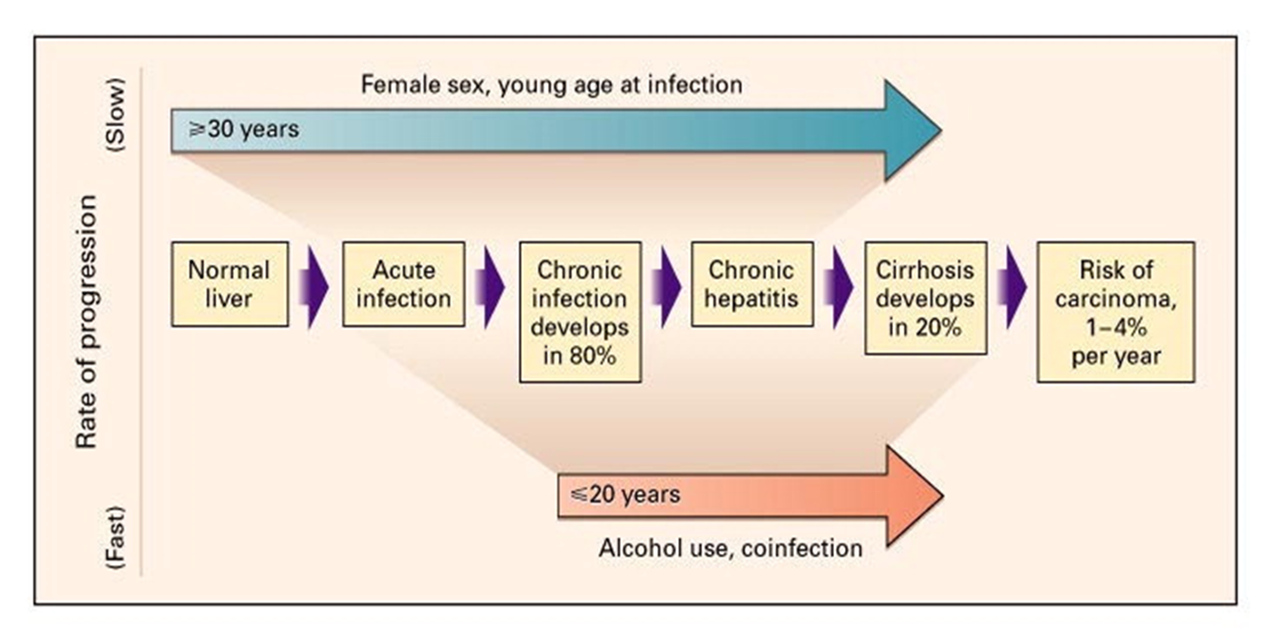
Although 15% to 25% of infected individuals spontaneously clear the virus without any treatment, approximately 75% to 85% will develop chronic HCV infection. The risk of cirrhosis after chronic HCV infection is 20% within 20 years and 30% within 30 years. Cirrhosis–related mortality from HCV ranges from 2% to 5% per year. Finally, in people with HCV cirrhosis, liver cancer occurs in an estimated 3% to 10% per year. Chronic HCV infection is the leading indication for liver transplantation in the U.S.1
Various epidemiologic risk factors can influence the risk and rate of HCV progression including age at acquisition, male sex, alcohol exposure, genetic factors, coinfection with other viruses, and other comorbidities (Figure 1).3,4
Fortunately, this life–threatening viral infection can now be eradicated in nearly all affected people. It is estimated that by 2025, HCV will be a rare disease.
Distinction between HCV as an infection, on the one hand, and a liver disease, on the other, is important. Most people with HCV have little or no visible evidence of liver damage, and if confirmed, management of the underlying infection is all that needs to concern the clinician. Once cured of HCV infection, no additional follow up is needed in this subset of patients. Those with significant liver damage need both antiviral therapy and attention to the potential consequences of a damaged liver.
HCV is a ribonucleic acid (RNA) virus classified in the Flaviviridae family. HCV replicates in the liver and in peripheral blood mononuclear cells, and it is detectable in serum during acute and chronic infection.5 The exact mechanism of viral entry into liver cells is not known, but it is associated with several viral and cellular factors. During HCV assembly and release from infected cells, virus particles associate with lipids and circulate in the blood in the form of triglyceride–rich particles.6 HCV polymerase is an enzyme encoded in the HCV genome that lacks error–correcting mechanisms, leading to a high mutation rate that likely contributes to the virus’ ability to evade the body’s immune system.7
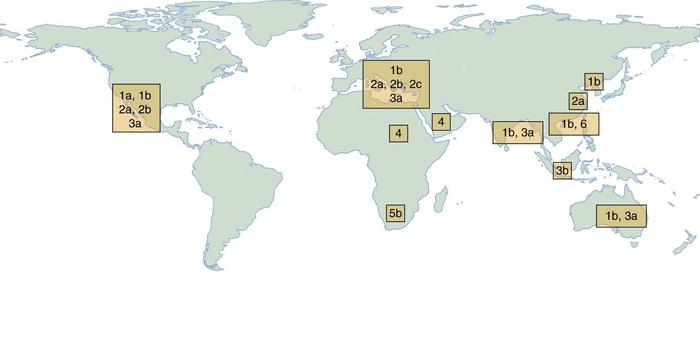
Genotype has considerable importance in determining the plan of treatment, although it does not affect the natural history of the disease. In the U.S., the predominant genotype is G1, constituting approximately 70% of individuals, 16% G2, 12% G3, and smaller proportions with genotypes 4, 5, or 6.8 Figure 2 indicates the geographic prevalence of HCV genotypes. Currently, G3 HCV is the most difficult to eradicate, although this may change with new treatments.
In the past, a major route of infection was via blood transfusion; however, effective screening of blood donations for HCV (since 1992) has reduced the risk of transfusion-associated HCV to less than 1 per 2 million units transfused.9 Currently, the most common route of transmission is related to intravenous drug use. Other modes of transmission include having multiple sexual partners, tattooing, body piercing, and sharing straws during intranasal cocaine use.10
Mother–child vertical transmission occurs in approximately 5% to 10% of cases and is more likely to occur in a mother who is coinfected with HIV.11 There are no conclusive data to advise against breastfeeding for women with HCV infection.12 Reports of infection rates after an HCV–contaminated needle–stick injury range from 0% to 10.3%, with an average rate of 0.5%, although not all infections lead to long–term infection.13
Transmission to a sexual partner in a monogamous relationship is uncommon with rates as low as 0.07% per year.14 Accordingly, the Centers for Disease Control and Prevention (CDC) does not suggest any change in sexual practice among monogamous couples.
The CDC and other public health experts recommend screening individuals with specific risk factors (Table 1).
| Screening for hepatitis C | CDC | USPSTF | VA |
|---|---|---|---|
| History of intravenous drug use | |||
| Blood transfusion before 1992 | |||
| Clotting factor use prior to 1987 | |||
| Long–term hemodialysis | |||
| Health care worker exposure | |||
| Chronic liver disease | |||
| Tattoo | |||
| Children of HCV mothers | |||
| Intranasal drug use (shared paraphernalia) | |||
| Alcoholic hepatitis, alcohol abuse, dependence | |||
| Age cohort 1945–1965 | |||
| Military service 1964–1975 |
Age Cohort Screening
Screening for hepatitis C is recommended for all individuals born between 1945 and 1965 ("Baby Boomers"). According to the CDC, nearly 75% of the 3.2 million Americans infected with chronic HCV were born between 1945 and 1965.17 This age cohort faces increased HCV–associated morbidity and mortality as roughly half of those infected with HCV are unaware of their HCV status and deaths from HCV are rising. According to national prevalence data, an adult in this age group is five times more likely than other adults to be infected with HCV, and as a whole this age group accounts for 73% of all HCV–associated deaths.18
One–time testing for antibodies to HCV (anti–HCV) by age–cohort was estimated to identify 800,000 more HVC infections. With subsequent treatment, testing by age cohort was estimated to avoid nearly 120,000 HCV–related deaths.18 Since implementation of age cohort testing, an estimated $1.5 to $1.7 billion dollars has been saved in liver disease–related costs.19
Acute HCV infection is uncommonly recognized because it is usually asymptomatic or accompanied by mild flu–like symptoms. Fatigue is the most common symptom. Other complaints include weight loss, muscle or joint pain, irritability, nausea, malaise, anorexia, depression, abdominal discomfort, difficulty concentrating, and jaundice. These symptoms occur 2 to 24 weeks after exposure.1
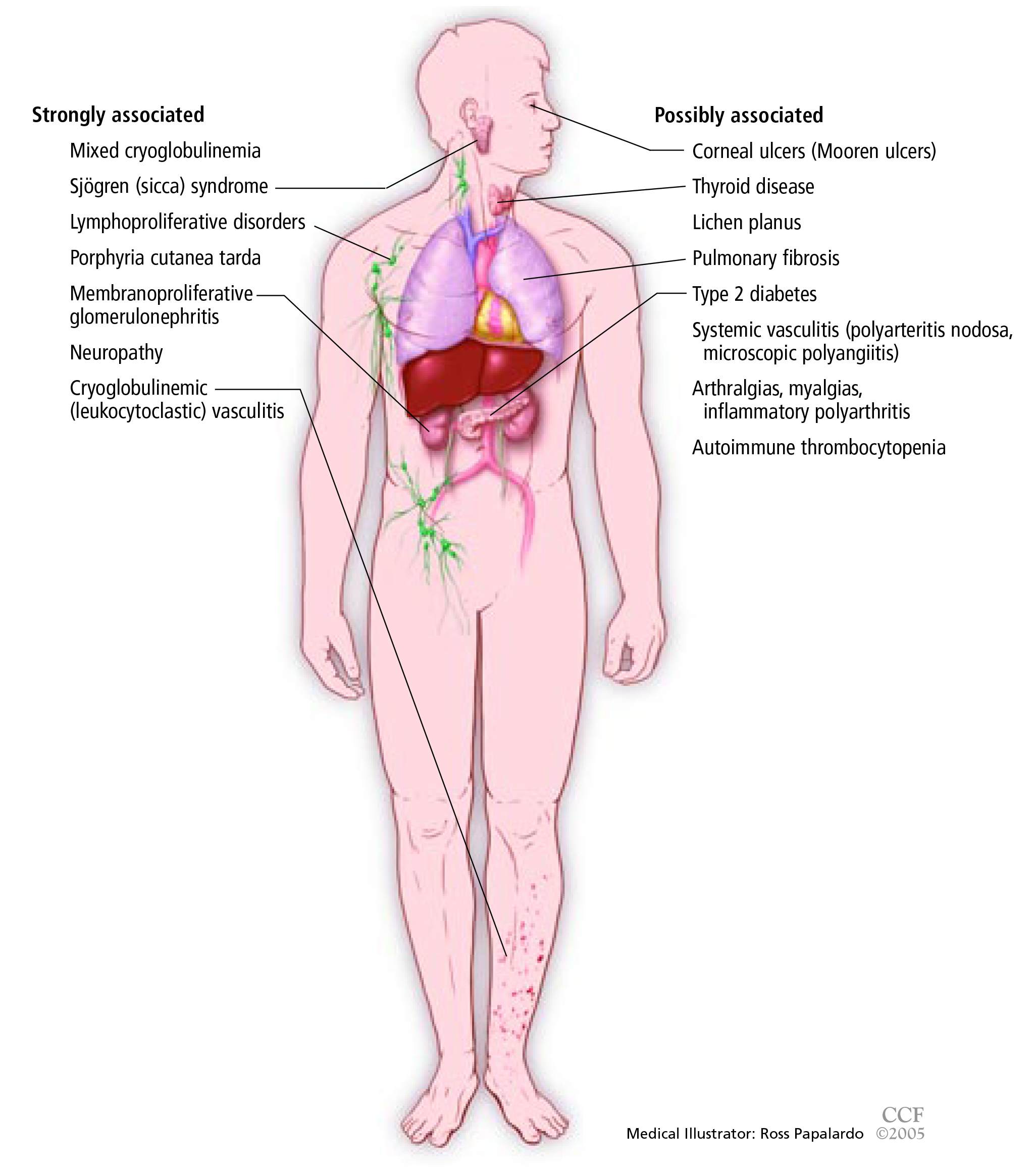
Many people with chronic HCV infection have no specific symptoms, and the finding of abnormal hepatic transaminase levels on routine testing often prompts specific testing for hepatitis C. Associated extrahepatic manifestations of HCV infection may include mixed cryoglobulinemia, arthralgias, porphyria cutanea tarda, and membranoproliferative glomerulonephritis (Figure 3).20, 21
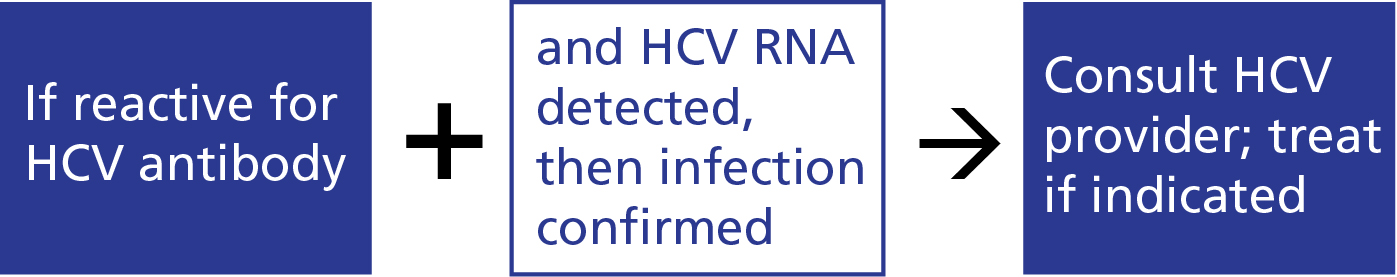
Diagnosis of HCV infection is usually a two–step process (Figure 4):
The most common serologic test used to detect HCV antibodies is the enzyme–linked immunosorbent assay (ELISA), which has a 95% sensitivity and specificity.22 A positive test, particularly if the person has a risk factor for transmission, almost always represents either active viral infection or resolved HCV. The assay can detect HCV antibodies 4 to 10 weeks after infection. Confirmation of active infection in any person with a positive anti–HCV assay requires the detection of HCV RNA by PCR. HCV RNA appears in the blood and can be detected 2 to 3 weeks after infection.22
There are two circumstances in which an individual infected with HCV may have a negative anti–HCV assay (a false negative result):
A high clinical suspicion is important if the first situation arises. Acute hepatitis C should be suspected if clinical signs and symptoms (eg, jaundice and ALT levels 10 times above the normal range) are compatible with infection. In most cases, HCV RNA can be detected during the acute phase and should be checked if clinical suspicion is high.25 In the second circumstance, an immunosuppressed patient may not be able to mount an antibody response. Again, if clinical suspicion is high, the HCV RNA level should be checked to confirm infection.
An HCV viral quantitative assay ("viral load") defines the presence and amount of virus. The viral load does not define the likelihood of disease progression, severity of disease, or degree of liver damage. The primary reason for checking HCV viral load is to have a baseline value prior to the initiation of treatment and then to monitor a person’s response to treatment after therapy is completed. It is not necessary to check the viral load repeatedly.
People infected with HCV may have normal or elevated ALT values, limiting the value of this test in the diagnosis of HCV. Although most HCV–infected individuals with persistently normal ALT values have no hepatic fibrosis, it may be present in up to 25% of this population.26
There is a surprisingly high incidence of advanced hepatic fibrosis in HCV–infected people. In one study of non–cirrhotic individuals with HCV infection, 56.2% had evidence of fibrosis on liver biopsy.27 Identifying people with advanced fibrosis (F3 or F4) is important as it determines prognosis and need for further monitoring. Individuals with advanced fibrosis need surveillance for hepatocellular carcinoma and routine blood work for the detection of progressive disease or complications related to their underlying cirrhosis.28 The role of hepatic fibrosis in HCV treatment decision–making is discussed later.
The absence of fibrosis also may have important implications in regards to prioritization and timing of treatment. The major barrier to treatment currently is the cost of therapy. Because the out–of–pocket cost for most people is too high, individuals are dependent on their insurance companies for access to medication. Generally, people with mild fibrosis (F0–F2) are less likely to be approved for therapy unless they have other HCV related comorbidities.29
Liver biopsy is an important method for determining the degree of fibrosis in certain situations and has been considered the gold standard. However, the procedure has flaws related to sampling variability and the potential for serious complications. In recent years, the role of liver biopsy in the assessment of liver fibrosis in HCV has diminished and given way to highly accurate noninvasive methods.
Of note, noninvasive markers may be very good at separating out the extremes, but are limited in distinguishing specific levels of fibrosis between the extremes. Thus, they are best for people with a very low probability of significant fibrosis or those with a high likelihood of cirrhosis.
When common laboratory tests, imaging, and physical exam findings point to cirrhosis, additional testing is often unnecessary. Absence of such findings, however, does not provide evidence for absence of hepatic fibrosis or even cirrhosis. Therefore, for many with HCV infection, additional assessment is advised. The two types of noninvasive tests most often used are serum–based tests and imaging studies.
There are both proprietary and nonproprietary serum–based tests to detect hepatic fibrosis. The nonproprietary models are just as useful as the proprietarily ones and are recommended. One commonly used nonproprietary serum marker model is the aspartate aminotransferase (AST) to platelet ratio index (APRI). In a meta–analysis of 40 studies, an APRI score ([AST level/upper limit of normal AST]/platelet count) greater than 1.0 had a 76% specificity and 72% sensitivity for predicting cirrhosis.30 An APRI score greater than 0.7 had a sensitivity of 77% and specificity of 72% for predicting significant hepatic fibrosis. Some evidence suggests that using multiple indices in combination or an algorithmic approach may result in higher diagnostic accuracy than using APRI alone. Another commonly used indirect serologic marker of fibrosis in Hepatitis C is the FibroSure assay, a proprietary assay that classifies the degree of fibrosis using the following variables: alpha–2 macroglobulin, haptoglobin, gamma globulin, apolipoprotein A1, GGT, total bilirubin, patient age, and sex. Results classify people from F0 to F4. This test has 60% to 75% sensitivity and 80% to 90% specificity for detecting significant hepatic fibrosis. Other indirect serologic markers of fibrosis include the HepaScore, AST/ALT ratio, FibroIndex, and ActiTest.31
Radiologic modalities are increasingly used for staging hepatic fibrosis. All are based on measuring the elastic property of liver. Hepatic elasticity is inversely related to the amount of fibrosis. FibroScan is one widely available device; ultrasound using acoustic radiation force impulse is another. Ultrasound–based elastography (also called transient elastography) measures hepatic tissue stiffness using shear waves. Ultrasound provides a rapid, inexpensive, and reproducible method of assessing fibrosis that is being increasingly utilized in the U.S. Magnetic resonance elastography is used less frequently.
Transient elastography evaluates a much larger area of the liver as compared with liver biopsy. The greatest experience with transient elastography has been staging hepatic fibrosis in people with hepatitis C. In one typical study using elastography, significant fibrosis was diagnosed consistently in hepatitis C.32 Elastography has an 87% sensitivity and 91% specificity for the diagnosis of cirrhosis. In 7 of 9 studies in a meta–analysis, transient elastography diagnosed stage F2 to F4 fibrosis with 70% sensitivity and 84% specificity.33
The major limitation of elastography is that results can be skewed in morbidly obese individuals. Elastography provides technically unreliable results in up to 15% of cases, and is especially unsuited for use in obese people.34 As in serum based testing, elastography is most reliable when used either in people with no fibrosis or cirrhosis.
We do not perform liver biopsies in most patients with HCV. According to the European Association for the Study of the Liver (EASL), noninvasive tests markedly reduce but do not abolish the need for liver biopsy.31
Liver biopsy is associated with significant risk. Nearly 84% report pain after liver biopsy.35 Although much rarer, some patients have even worse complications: Severe bleeding occurs in 0.35% to 0.5%, perforation in 0.57%, and even death in approximately 0.03%.35 Not only do patients find liver biopsy anxiety–provoking, but physicians are averse to performing it. A recent survey of 104 physicians found that only 46.2% of them perform liver biopsy as the primary diagnostic tool.36
In addition, the amount of tissue obtained by a needle liver biopsy represents 1/50,000 of the total liver volume.37 As hepatic fibrosis is not uniformly distributed, there can be many false results. In one study, 124 people with chronic HCV underwent biopsy of their left and right hepatic lobes. The biopsy samples showed a difference in the intensity of inflammation in 24.2% of cases and in the intensity of fibrosis in 33.1% of cases. In 14.1% of cases, cirrhosis was diagnosed in one lobe but not the other.38
Considered through the prism of individual and public health, even considering total societal costs, the most effective strategy is clearly to treat all HCV–infected people despite the high cost of medication.39–41 All individuals at any stage of hepatitis C should be considered for treatment. Higher overall cure rates improve patient–related outcomes, increase worker productivity, and ultimately lessen the economic burden of chronic HCV on society.42, 43
Chronic HCV–related liver disease puts a tremendous economic burden on infected individuals, their families, and society as a whole. In addition, as chronic HCV is a systemic disease with the potential to affect not only the liver but other organ systems, the disease burden can be enormous. From an economic standpoint, successfully treating this disease positively affects the entire community.42
Moreover, those cured of chronic HCV have a reduction in the rate of progression of liver fibrosis and an overall decrease in liver inflammation. This, in turn, is associated with a more than 70% reduction in the risk of hepatocellular carcinoma and 90% reduction in the risk of liver–related death and liver transplantation.44,45 Achieving cure in the earlier stages of fibrosis (F0 or F1) can prevent many of the complications of advanced liver disease (F3 or F4) such as hepatic decompensation or hepatocellular carcinoma.44,45
Achieving cure has been shown to improve social, physical, and emotional health of those infected with HCV. In a large study of 4,781 people with chronic HCV infection, both depression (found in 30%) and poor physical health (found in 25%) were associated with unemployment, higher stressful events, and lower social support. Cure of HCV was associated with a lower risk of depression and with better physical health.46
Benefits of cure accrue regardless of the stage of fibrosis at baseline. Even people with advanced fibrosis or cirrhosis realize as much as an 80% reduction in the risk of developing clinical decompensation.47 A number of studies have also shown a decrease in the risk of specific complications, including a decrease of as much as 77% in the risk of development of hepatocellular carcinoma.44 Fibrosis may also improve. Among those with lesser degrees of liver damage, studies suggest a decrease in the degree of liver fibrosis in treated individuals and, in a minority of people, regression of cirrhosis.48
All treatments for Hepatitis C are very expensive. Failure to adopt a treat–all approach derives mainly from the high cost of medication, and the divided fiscal responsibilities and loyalties within the healthcare system. Companies and agencies that underwrite prescription benefits have not factored in to their premiums the explosion of very expensive medications for HCV. The greatest current barrier to treating HCV is the financial burden of the very expensive therapies.49 For example, as of September 2016, a 12–week course of sofosbuvir–velpatasvir (Epclusa) costs $89,712, or $1,068 per pill.50 Sofosbuvir–ledipasvir (Harvoni) costs $1,350 per pill.51 As most people cannot cover these costs, payment and authorization for treatment falls to insurance companies.
Because of limited resources, insurers have established a triage strategy that aims to provide treatment for individuals with more advanced disease. Preauthorization for payment for treatment is most often based on fibrosis stage (F3 or F4 fibrosis), risk of progression towards more advanced disease, increased risk of HCV transmission, and extra–hepatic manifestations of HCV. Other factors may include people with HIV or HBV coinfection, or decompensated cirrhosis. Some insurers also take into account the presence of comorbidities such as lymphoma, renal disease, hepatocellular carcinoma, and transplant patients. Most require demonstration of freedom from drug use (urine drug toxicology screening) before they will agree to pay. Other less critical factors considered in approval of treatment include difficulties with compliance (eg, missed doctor’s appointments), inadequate social support, mental health disorders, and having a history of substance abuse.
There have been recent court challenges against insurers for payments for HCV therapy. The effect of this combined with the introduction of additional less expensive treatments will likely make it easier and less costly for HCV–infected individuals to get therapy.
The goal of treatment is permanent elimination of virus. Absence of virus in the blood 12 weeks after ending treatment is referred to as sustained virologic response (SVR). Increasingly, SVR and “cure” are used interchangeably and will be so used in this chapter. By convention, SVR determination is made 12 (or more) weeks after cessation of antiviral treatment. This is sometimes called SVR 12. Those who achieve an SVR have HCV antibodies but no longer have detectable levels of HCV RNA.
The ideal treatment for HCV would have the following attributes:
Currently available treatments achieve all of these goals except for low cost. Put another way, the only barrier to elimination of HCV is the high cost of medication. The most widely used medications combine in a single pill two drugs attacking different steps of HCV viral replication and assembly. In a few circumstances, it is necessary to add a second pill, ribavirin. Wherever possible we avoid using this agent because it adds complexity and side effects (principally anemia), and because it cannot be safely used in women who are or may become pregnant or in men whose partners may become pregnant.
Pretreatment assessment of the patient with chronic hepatitis C should include the following:
Currently, there are 10 FDA–approved direct–acting agents marketed by 8 different companies for HCV treatment. Combining two or more agents targeting different steps in the viral replication process has proven to be the most successful strategy. This chapter focuses on those regimens that make the most sense for general practitioners in the U.S. to treat people without decompensated cirrhosis, advanced renal insufficiency (eGFR >30), or coinfection with hepatitis B or HIV. Individuals with advanced cirrhosis or with a coinfection (HIV or HBV) should be seen by a healthcare provider with special expertise in gastroenterology, hepatology, or infectious disease. Those with significant renal insufficiency (eGFR <30) also should be treated by a specialist.
Many HCV treatment regimens use sofosbuvir, a nucleotide pro–drug. The activated form of sofosbuvir acts as a defective substrate for the RNA–dependent viral nonstructural protein 5B (NS5B) RNA polymerase and is incorporated into viral RNA and terminates synthesis, resulting in disruption of viral replication. This, in turn, leads to viral death.
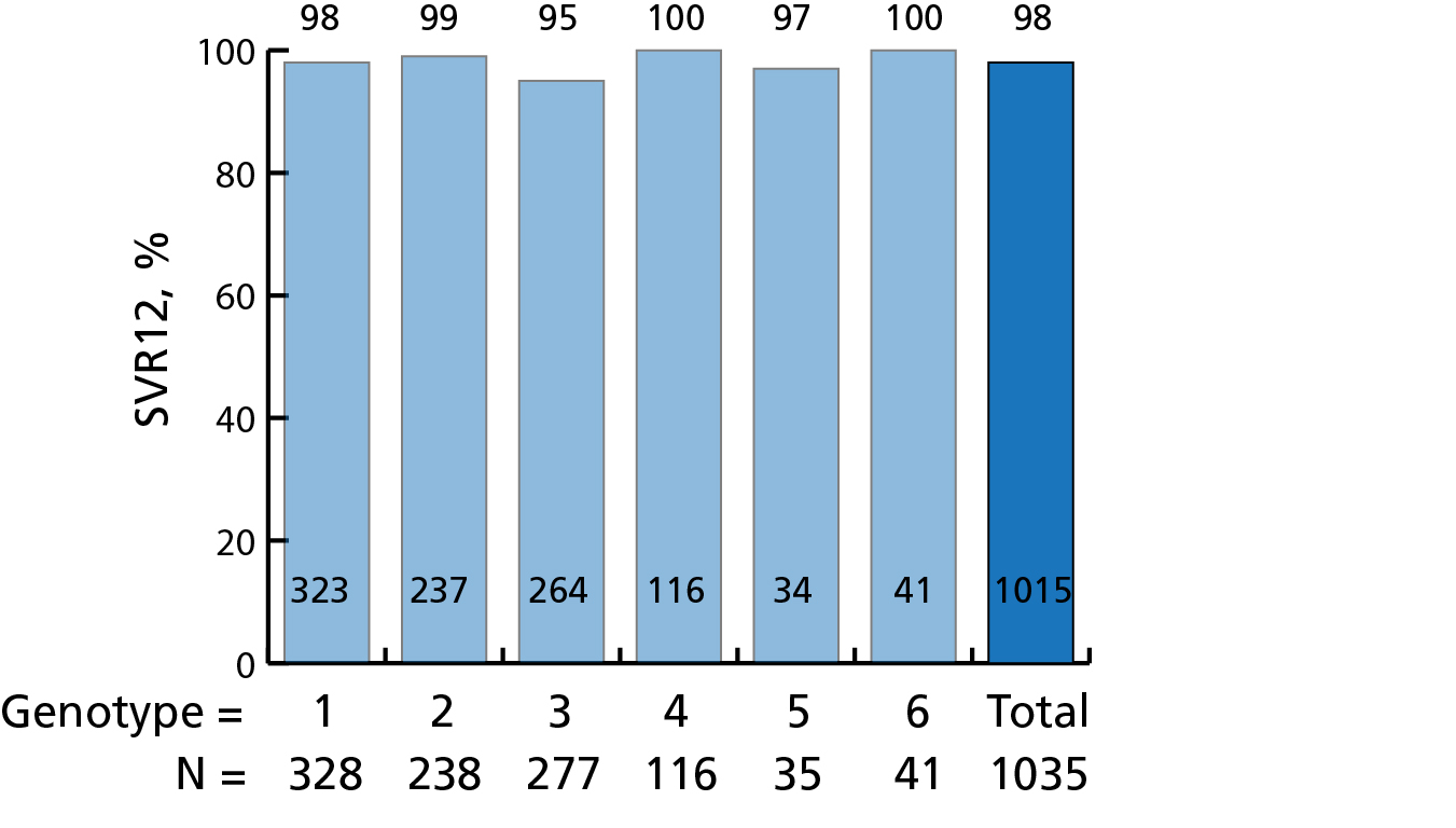
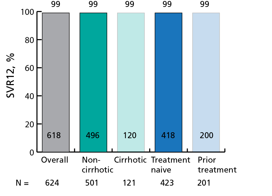
Administration of sofosbuvir, an NS5B polymerase inhibitor, combined with either the NS5A inhibitors velpatasvir (Epclusa, a once–daily, fixed–dose combination tablet) or ledipasvir (Harvoni) results in cure rates of 95% to 100% after 12 to 24 weeks of treatment.28 Velpatasvir–sofosbuvir is particularly attractive because it can be used for all HCV genotypes
(Figure 5 and Figure 6).52, 53 Either drug combination can be used regardless of whether the patient has cirrhosis. Therefore, a person with chronic HCV without cirrhosis or with compensated cirrhosis (defined as Child–Pugh Class A) of any HCV genotype could be treated with velpatasvir–sofosbuvir. If the genotype is 1, 4, 5, or 6, ledipasvir–sofosbuvir is an equivalent choice (Table 2). A person with decompensated cirrhosis (defined as Child–Pugh Class B or C) of any HCV genotype should be referred to a specialist.
| Polymerase inhibitors | ||||
| Protease inhibitors |
Nucleotide Nonnucleoside | NS5A protein inhibitors |
Brand name(s) |
Target genotype(s) |
| Sofosbuvir | Velpatasvir | Epclusa | 1,2,3,4,5,6 | |
| Sofosbuvir | Ledipasvir | Harvoni | 1,4,5,6 |
Drug regimens listed in this section are highly effective but are more restricted by the hepatitis C genotypes (Table 3). Some require an additional medication such as ribavirin, a drug that has additional side effects and toxicities, especially anemia and teratogenicity.
| Polymerase inhibitors | ||||
| Protease inhibitors |
Nucleotide Nonnucleoside | NS5A protein inhibitors |
Brand name(s) |
Target genotype(s) |
| Paritaprevir/ritonavir | Dasabuvir | Ombitasvir | Viekira XR | 1a*, 1b |
| Grazoprevir | Elbasvir | Zepatier | 1a†, 1b†, 4† | |
| Sofosbuvir | Daclatasvir | Sovaldi, Daklinza | 3‡ | |
| Paritaprevir/ritonavir | Ombitasvir | Technivie | 4‡‡ | |
*For HCV genotype 1a without cirrhosis, add ribavirin and treat for 12 weeks; for genotype 1a with compensated cirrhosis, add ribavirin and treat for 24 weeks.
†For HCV genotype 1a without NS5A polymorphism, or for genotype 1b, or for genotype 4 not previously treated, treat for 12 weeks; for genotype 1a with NS5A polymorphism, or for genotype 4 that was previously treated, add ribavirin and treat for 16 weeks.
‡For HCV genotype 3, treat with Daklinza with Sovaldi for 12 weeks; longer if patient has cirrhosis.
‡‡For HCV genotype 4 without cirrhosis, treat with Technivie plus ribavirin for 12 weeks; Technivie without ribavirin for 12 weeks may be considered for treatment–naïve patients who cannot take/tolerate ribavirin.
The combination of dasabuvir, ombitasvir, paritaprevir, and ritonavir (Viekira XR/Viekira Pak) is a highly effective regimen for people with genotype 1 HCV. Viekira XR differs from Viekira Pak. Viekira XR is a once–daily, extended release combination pill containing all of the active ingredients found in the Viekira Pak. Viekira XR consists of 3 pills taken once daily versus the twice daily dosing of Viekira Pak.
The single–pill combination of elbasvir and grazoprevir (Zepatier) is taken once daily. It is highly effective for those with genotype 1 or 4. Two questions must be answered to tailor its use to optimal effect:
Elbasvir–grazoprevir may be used as monotherapy for 12 weeks in those with the following:
If the HCV infection is genotype 1a and baseline NS5A polymorphism is present, coadministration of ribavirin with elbasvir–grazoprevir is recommended. Similarly, 16 weeks of both drugs is given in HCV genotype 4 if interferon and ribavirin have been used previously.
Miscellaneous agents and combinations include daclatasvir that can be used for 12 weeks along with sofosbuvir in HCV genotype 3. It is highly effective (92% to 98% cure rate) when there is no cirrhosis but has unacceptably low success (58% to 69% cure rate) when cirrhosis is present. Sofosbuvir and simeprevir has been used in genotypes 1a and genotype 1b in people with cirrhosis.
The direct–acting antivirals have an excellent side effect profile and are generally well tolerated. The most common side effects of the Epclusa (velpatasvir–sofosbuvir) combination pill (at 12 weeks) are headache (22%), fatigue (15%), nausea (9%), asthenia (5%), and insomnia (5%).52
The most common side effects of the (Harvoni) ledipasvir-sofosbuvir combination pill (at 12 weeks) are fatigue (13%), headache (14%), nausea (7%), diarrhea (3%), and insomnia (5%).54
A major drug interaction with both of these drugs occurs with amiodarone. Serious symptomatic bradycardia (slow heart rate) has been associated with velpatasvir–sofosbuvir and amiodarone, especially in those with advanced liver disease or underlying heart disease. Therefore, coadministration of velpatasvir–sofosbuvir or amiodarone is not recommended for people without alternative treatment options.
In addition, drugs that are inducers of P–glycoprotein and/or inducers of certain cytochrome P enzymes may decrease the serum concentrations of sofosbuvir and/or velpatasvir resulting in a decreased potency of the combination. Certain established clinically significant drug interactions have also been described.
As with any medication regimen, individuals should be monitored while on hepatitis C therapy to ensure medication compliance, assessment for potential adverse reactions, and avoids potential drug–drug interactions.
Confirmation of treatment success is demonstrated by the absence of HCV virus in the blood 12 or more weeks after completion of therapy. Such a finding represents a cure. Those with stage F0, F1, or F2 fibrosis who have achieved SVR no longer need to be monitored for their liver disease. However, the individuals who have F3 or F4 fibrosis and achieve SVR should be monitored for cirrhosis. They also should be screened for hepatocellular carcinoma every 6 months with a right upper quadrant ultrasound. In addition, most require an upper endoscopy for screening for varices, usually by a gastroenterologist or a hepatologist.
Individuals who fail to achieve an SVR should continue to have routine lab work (ie, complete blood count, hepatic function panel, international normalization ratio) every 6 months to monitor their liver function.25, 28 Evaluation for retreatment by a hepatologist or gastroenterologist is recommended if initial treatment fails.
The only contraindication to current chronic HCV treatment is in a patient with a short–life expectancy that cannot be lengthened with treatment, with liver transplant, or with any other treatments.
Acute HCV infection is frequently undiagnosed due to lack of specific symptoms. Because up to 50% of acutely infected individuals will spontaneously clear the infection, a balance must be struck between watchful waiting and treatment. At minimum, monitoring for HCV RNA for at least 3 months before starting treatment is recommended to allow for spontaneous clearance; however, delaying for 6 months is also acceptable. In people who are anti–HCV positive and HCV RNA negative (ie, spontaneous clearance of infection), HCV RNA should be tested again in 3 months to confirm true clearance.
Healthcare workers accidentally exposed to HCV-infected blood via a needle–stick injury should immediately report the exposure. They should have immediate testing for anti–HCV antibodies to establish the absence of pre–existing infection. There is no value to administration of either serum immune globulin or prophylactic antiviral treatment. The average incidence of anti–HCV seroconversion after needle–stick from a known anti–HCV positive source is 0% to 10.3%.13
For individuals diagnosed with acute HCV infection (ie, positive anti–HCV antibodies and positive HCV RNA), there is no clear benefit to early treatment. Monitoring for 6 months is acceptable as most cases will spontaneously clear without intervention. When the decision is made to initiate treatment after 6 months, treating as described for chronic hepatitis C is recommended, given its high efficacy and safety.28 In addition, insurance reimbursement plans have not made an exception to the general requirement that significant fibrosis must be present for payment, making the issue of early treatment essentially moot.
Studies of the predictors of spontaneous clearance of HCV infection have suggested that clearance may be more likely to occur in younger people and in those with a more symptomatic presentation, particularly with jaundice.55
These individuals are best managed by specialists, so they are only briefly described here.
Decompensated cirrhosis is defined as Child–Pugh Classification B or C. It also can be defined clinically by the development of liver decompensation, such as jaundice, or complications of portal hypertension, such as ascites, variceal hemorrhage, or hepatic encephalopathy Treatment decisions for people with decompensated cirrhosis should be made by a hepatologist, preferably in the context of a liver transplant program.
Infection of the new liver is almost universal posttransplant, and the subsequent rate of hepatic fibrosis can be quite rapid. The likelihood of a patient developing cirrhosis in the newly transplanted liver over the course of 3 to 5 years posttransplant, is as high as 10% to 30%.56
In a small subgroup of individuals following liver transplant with HCV (27 of 179), the pattern of recurrence, with mainly cholestatic features, and the rate of disease progression is much more aggressive, leading to death in 1 to 3 years in up to 60% of these individuals.57
This same group at high risk of progression has also traditionally been more difficult to treat successfully. Patients with HCV recurrence post liver transplantation tend to have elevated viral loads – on average 10– to 20–fold greater than pretransplant levels.58 Several treatment regimens, most commonly involving sofosbuvir–ledipasvir, can be used in post transplant individuals with HCV depending on the specific genotype.28 The exact treatment regimen, however, should be determined by a hepatologist.
There are no reports of the safety or efficacy of velpatasvir–sofosbuvir in liver–transplant recipients. However, neither sofosbuvir nor velpatasvir interacts with cyclosporine or tacrolimus, the most commonly used immunosuppressants after liver transplant. Based on the lack of real–world experience in liver–transplant recipients, velpatasvir–sofosbuvir should not be used for this particular population.28
Hepatitis C adversely affects survival in those with chronic kidney disease, especially those on dialysis. In addition, HCV seems to play a role in the rate of progression of kidney disease. Drug metabolism of many of the drugs used for HCV is altered in people with severe renal impairment and those on dialysis. For all of the drug regimens mentioned in this chapter except elbasvir–grazoprevir, data on dosage adjustments for those with eGFR <30 are not available. For elbasvir–grazoprevir (Zepatier), standard doses are recommended at all levels of renal insufficiency and for those on dialysis.
Cure rates with currently available therapies are often disappointing. Elbasvir–grazoprevir has been shown to cure 94% of those with HCV genotype 1 and 4 and advanced renal disease, including those on dialysis.59 Other trials are currently ongoing. For those with genotype 1 or 4, 12 weeks of elbasvir–grazoprevir is recommended. The combination of dasabuvir, ombitasvir, paritaprevir, and ritonavir is an alternative in those with genotype 1b. Currently, those with severe renal insufficiency/dialysis and genotype 2, 3, 5, or 6 are either not treated or given pegylated interferon and dose–adjusted ribavirin.
Patients with HCV infection often share risk factors for coinfection with both hepatitis B virus (HBV) and HIV. HIV is known to accelerate HCV disease progression. With the use of direct–acting antivirals, those coinfected with HIV and HCV can be reliably treated with a traditional 12-week course.
The importance of treatment is clear – those successfully treated are at a substantially lower risk of developing liver disease, complications of liver disease, or suffering a liver–related death.60 However, there are major issues with drug–drug interactions with sofosbuvir–velpatasvir and several of the medications used to control HIV, the highly active antiretroviral therapies, that may affect the efficacy of treatment (Table 4).
| HIV antiviral | Interaction | Recommendation |
|---|---|---|
| Efavirenz | Decreases the levels of velpatasvir | Coadministration is not recommended |
| Regimens containing tenofovir | Increases the levels of tenofovir | Monitor closely for adverse reactions to tenofovir when administered with sofosbuvir–velpatasvir |
| Tipranavir and ritonavir | Decrease the levels of sofosbuvir and velpatasvir | Coadministration is not recommended |
The clinical course of HCV is thought to be worsened by HBV coinfection. Those with chronic HCV and active HBV coinfection have a more severe degree of liver fibrosis and a higher risk of developing hepatocellular carcinoma than with HCV alone. Some studies, however, have suggested that an active HBV infection, but not previous infection, actually suppresses the replication of H. One study in anti–HCV positive subjects found a significantly lower prevalence of HCV RNA (41%) in those with active HBV infection than in those who had recovered from HBV infection (82%).61
Several cases of reactivation of dormant hepatitis B have been reported when treatment of HCV is undertaken, including a few with severe liver injury requiring urgent liver transplantation.62 Those who are anti–HBc positive (whether they have HBsAg and even when HBV DNA levels are undetectable) are at risk. Because of this, we recommend referral of these people to a specialist for HCV treatment whenever anti–HBc or HBsAg is present.
Although there are data on the use of interferon, treatment for HBV–HCV coinfection has not been thoroughly studied regarding direct–acting antiviral agents. These individuals should be treated by a hepatologist.
Currently, alcohol use and HCV infection are the two primary causes of cirrhosis and liver transplantation in the U.S. Concurrent alcohol use is a common occurrence in people with chronic HCV. Because alcohol intake varies over time and affects individuals differently, it has been difficult to find an exact threshold for the risk to a person with HCV infection and concomitant alcohol use.
It is clear, however, that heavy alcohol use (defined as 5 or more drinks per day) contributes to HCV–associated liver disease. In a study of 6,600 people with HCV, higher alcohol intake was associated with significantly more cirrhosis compared with lower alcohol intake. Moreover, studies have shown that even lower amounts of alcohol were associated with fibrosis, although to a lesser extent than heavy alcohol use.63, 64
Therefore, it is quite clear that alcohol intake increases the risk of cirrhosis alone and particularly in combination with HCV. As cirrhosis is the primary risk factor for hepatocellular carcinoma, alcohol use is associated with this cancer risk through its cirrhosis effect. Alcohol itself does not interact with the velpatasvir and sofosbuvir or other direct–acting antivirals.
According to the U.S. Department of Veteran’s Affairs, those with chronic HCV likely should not drink alcohol as any amount can contribute to worsening liver disease.63 Heavy alcohol use, in particular, has been linked with fibrosis progression and cirrhosis although even light and moderate amounts do so to a lesser degree. People with established cirrhosis should definitely abstain from alcohol use.
Women who are pregnant are not routinely tested for hepatitis C unless they are at risk (eg, IV drug users). Women who test positive for hepatitis C do not need to be treated while pregnant. The average rate of HCV transmission from a HCV–infected mother to child is 5% to 6% if the mother is HIV–negative and up to 14% if the mother is HIV–positive.65 There is no known method of preventing perinatal transmission of HCV. Luckily, spontaneous resolution of HCV occurs in approximately 50% of HCV–infected infants within the first 3 years of life, so HCV treatment should not be considered before 3 years of age.65 Children who do not clear the virus should be closely followed by a specialist.
Hepatitis C virus is not spread through breast milk. The Centers for Disease Control endorses breast feeding in HCV infected women unless her nipples are cracked and bleeding, or in the event of HIV–HCV coinfection.66
Occasionally, a woman being treated for HCV will become pregnant. Currently, there are no adequate human data available to establish whether sofosbuvir plus velpatasvir poses a risk to pregnancy outcomes. In addition, it is not known whether the components of sofosbuvir–velpatasvir and their breakdown products are present in human breast milk, affect human milk production, or have effects on breastfed children.
Any combination that contains ribavirin, a known teratogenic agent, must be assiduously avoided in both female and male partners. At least 2 reliable methods of birth control are recommended for any individual who is receiving ribavirin or who has done so within the preceding 6 months. The risks-benefits of breastfeeding if considering HCV treatment with sofosbuvir–velpatasvir should be discussed with a specialist prior to initiating treatment.
Not surprisingly, given the prevalence of HCV infection, many individuals, perhaps 30% to 40%, have explored complementary and alternative medicines (CAM) often without the knowledge of their healthcare providers.67 The usefulness of most CAM therapies, including dietary supplements, herbs, and unconventional treatments, has not been rigorously studied, and the results are extremely varied. Many take milk thistle, whose active ingredient silymarin is thought to function as an antioxidant. Although it has been shown in lesser quality studies to have multiple effects on the virus lifecycle, including inhibition of specific viral enzymes and cell entry, clinical trials have not shown consistent benefit.68 A 2005 Cochrane meta–analysis reviewing 13 randomized clinical trials demonstrated that milk thistle, although very safe to use, had no significant effects on mortality, complications of liver disease, or liver histology.69 Other CAM options that have been studied, albeit in studies of varying degrees of quality and with variable sample sizes, have tested vitamin E, thymus extract, zinc, traditional Chinese medicine, glycoprotein, glabra (licorice root), and oxymatrine. Most interventions have only marginal benefit, and the National Center for Complementary and Alternative Medicine review on the use of herbal treatment concludes that no CAM treatment has been proven to successfully treat HCV.70
Recent studies also have examined the role of diet and response to treatment or progression of disease. Although an extremely common issue, there are limited data to suggest a role for specific dietary modification, although it may be reasonable to minimize intake of polyunsaturated fatty acids, fats, and carbohydrates during treatment.71
HCV infection typically becomes chronic in more than 60% of cases and it can lead to cirrhosis in as many as 20% over a 20–year period. Serum aminotransferase levels reflecting hepatocellular injury can fluctuate, as does the viral load, making them unreliable markers of disease severity.
As the disease evolves, hepatocytes are progressively destroyed and replaced by fibrosis, slowly leading to the development cirrhosis unless it is treated. The pathologic course is affected by various factors, such as the patient’s age at onset of infection, sex, coinfection with other viruses, other medical conditions, and risk behaviors. The risk of developing hepatocellular carcinoma in people with chronic HCV plus cirrhosis is as high as 4% per year. HCV–infected individuals with cirrhosis should be followed by a liver specialist.
Direct–acting antiviral agents have made it possible to cure most cases of HCV infection. Although successful treatment with these regimens is associated with a lower rate of liver–related complications, and perhaps even regression of fibrosis/cirrhosis, those successfully treated may still be at risk for hepatocellular carcinoma if they have cirrhosis.
Although HCV remains a major global health problem, significant advances in the understanding of its basic biology have allowed improvements in treatment. Given the pace of discovery in this field, it may one day be possible to eradicate this deadly virus.
Neal Mehta, MD; nothing to disclose. William Carey, MD; nothing to disclose. Naim Alkhouri, MD; nothing to disclose. Robert S. O’Shea, MD, MSCE; nothing to disclose.