Cleveland Clinic physicians present selected topics about the management of common diseases and conditions.
Syncope
Erika Hutt Centeno, MD; Kenneth A. Mayuga, MD; Fetnat Fouad-Tarazi, MD; Laura Shoemaker, DO; Fredrick Jaeger, DO
Atopic Dermatitis
Melissa Piliang, MD; Sarah Schneider, MD
Melanoma
Alok Vij, MD
Nonmelanoma Skin Cancer
Daniel Knabel, MD; Allison Vidimos, MD
Diabetes Mellitus Treatment
Mario Skugor, MD
Erectile Dysfunction
Milton Lakin, MD; Hadley Wood, MD
Hypercalcemia
Mario Skugor, MD
Colonic Diverticular Disease
Vladimir Bolshinsky MD; Scott R. Steele, MD, MBA; Prashanthi N. Thota, MD
Acute Liver Failure
Arvind R Murali, MD; KV Narayanan Menon, MD
Alcoholic Liver Disease
Catherine Frakes Vozzo, DO; Nicole Welch, MD; Carlos Romero-Marrero, MD; Kyrsten D. Fairbanks, MD
Alpha1-Antitrypsin Deficiency
Anthony S. Tavill, MD; Loutfi S. Aboussouan, MD
Autoimmune Hepatitis
Andrea Fialho, MD; Andre Fialho, MD; William D. Carey, MD
Cirrhotic Ascites
Karin B. Cesario, MD; Anuja Choure, MD; William D. Carey, MD
Gallbladder and Biliary Tract Disease
David S. Barnes, MD
Hemochromatosis
Anthony S. Tavill, MD
Hepatic Encephalopathy
Mina Shaker, MD; William D. Carey, MD
Hepatitis A
Talal Adhami, MD; Ibrahim Hanouneh, MD
Hepatitis B
Robert S. O'Shea, MD
Hepatitis C
Neal Mehta, MD; William Carey, MD; Naim Alkhouri, MD; Robert S. O'Shea, MD
Hepatitis D
Talal Adhami, MD; William D. Carey, MD
Hepatitis E - An Evolving Disease
Sandra Rodriguez, MD; William D. Carey, MD
Liver Disease in Pregnancy
Jamilé Wakim-Fleming, MD
Liver Test Interpretation - Approach to the Patient with Liver Disease: A Guide to Commonly Used Liver Tests
Arvind R. Murali, MD; William D. Carey, MD
Nonalcoholic Fatty Liver Disease
Emily Carey, DO; Anna Wieckowska, MD;
William D. Carey, MD
Post-Liver Transplantation Management
Bijan Eghtesad, MD; Charles M. Miller, MD;
John J. Fung, MD
Primary Biliary Cirrhosis, Primary Sclerosing Cholangitis, and Other Cholestatic Liver Diseases
Claudia O. Zein, MD
Variceal Hemorrhage
Karin B. Cesario, MD; Anuja Choure, MD; Kunjam Modha, MD; William D. Carey, MD
Wilson's Disease
Anthony S. Tavill, MD
Adult Immunization
Patricia Dandache, MD
Sepsis
Bethany Lehman, DO; Patricia Dandache, MD
Headache
Jennifer S. Kriegler, MD
Multiple Sclerosis
Carrie M. Hersh, DO, MSc; Robert J. Fox, MD
Paget's Disease of Bone
Elizabeth File, MD; Abby Abelson, MD
Breast Disorders, Cancer Screening, and Risk Assessment
Holly J. Pederson, MD; Shazia Goraya, MD
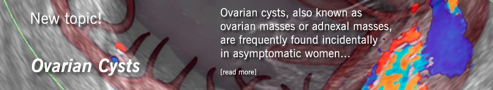
Endometrial, Ovarian, and Cervical Cancer
Elizabeth Connor, MD; Chad Michener, MD
A generous gift in memory of Eileen Cawley made this work possible.
Take Disease Management Clinical Decisions lessons for CME credit