Published: July 2019
Expire: July 2022
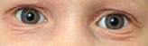
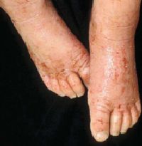
Atopic dermatitis (Figure 1) and atopic eczema1 are interchangeable terms for an inflammatory condition of the skin characterized by erythema, pruritus, scaling, lichenification, and papulovesicles. Atopic dermatitis is a distinct condition in persons who are genetically predisposed to developing immunoglobulin (Ig) E-mediated hypersensitivity reactions. It is characterized by the itch-scratch cycle: affected persons have the sensation of itch, followed by scratching and the subsequent creation of a rash. The classic triad of atopy includes atopic eczema, asthma, and allergies. A wide range of environmental factors such as contact allergens, stress, food, skin flora, and humidity play roles in the development and severity of atopic dermatitis.
Atopic dermatitis is a common condition affecting approximately 17% of the population2 with a slight female preponderance (1.3:1 in children). The incidence has increased twofold to threefold since the 1970s. The basis of this increase is not well understood; however, environmental factors appear to play an important role in disease prevalence.
Factors associated with an increased risk of atopic dermatitis include small family size, higher socioeconomic and educational levels regardless of ethnicity, movement from a rural to urban environment, and increased use of antibiotics (the Western lifestyle). This has led to the hygiene hypothesis, which suggests that infections in early childhood (from less-hygienic practices and older siblings) might prevent atopic dermatitis. This hypothesis is supported by evidence that infections induce type-1 helper T cells (TH1), whereas there is a predominance of type-2 helper T cells (TH2) in atopic dermatitis. TH1 responses antagonize the development of TH2 cells, thereby potentially decreasing the incidence of atopic dermatitis.3
Atopic dermatitis is a type I IgE-mediated hypersensitivity reaction, but the exact etiology is unknown. The pathogenesis is multifactorial and involves a complex immunologic cascade, including disruption of the epidermal barrier, IgE dysregulation, defects in the cutaneous cell-mediated immune response, and genetic factors.
The major elements in immune dysregulation are Langerhans' cells, inflammatory dendritic epidermal cells, monocytes, macrophages, lymphocytes, mast cells, and keratinocytes, all of which interact through an intricate cascade of cytokines leading to a predominance of TH2 cells over TH1 cells.4 The TH2 cytokines, interleukin (IL)-4, IL-5, IL-10, and IL-13, are increased in the skin, and there is a corresponding decrease in TH1 cytokines, mainly interferon-gamma and IL-2.
Patients with atopic dermatitis often have dry, sensitive skin due to changes in the epidermis, which serves as a barrier to the environment by maintaining the water balance of the skin. Essential fatty acids (EFAs), such as linoleic and linolenic acid, are important components of the epidermal barrier. In atopic dermatitis, delta-desaturase activity is deficient,5 which leads to decreased linoleic and linolenic acid metabolites. Loss of EFAs results in increased transepidermal water loss and subsequent xerosis (dryness). The EFAs form the substrate of the inflammatory mediators (prostaglandins and leukotrienes), resulting in a secondary deficiency of prostaglandin E1.
Defects in the epidermal barrier also lead to increased susceptibility to atopens (atopic allergens such as house dust mites, grass, or pollen). When such allergens contact atopic skin, they stimulate TH2 lymphocytes to produce cytokines such as IL-4, IL-5, and IL-13, which in turn promote an increase in IgE synthesis.6 Atopic dermatitis patients often have high levels of IgE antibodies due to house dust mites and other allergens. Eliminating these allergens from the environment, an extremely difficult undertaking, can improve atopic dermatitis.
Defective cell-mediated immunity leads to increased susceptibility to many bacterial, viral, and fungal infections of the skin. Children with atopic dermatitis are particularly susceptible to severe, widespread herpes simplex virus infection (eczema herpeticum)—a systemic and potentially fatal infection that primarily affects areas of active atopic eczema. Widespread infections with human papillomavirus (warts) and molluscum contagiosum are also common in children with atopic dermatitis.
In Dermatology, the microbiome includes resident microorganisms of the skin and plays an important role in regulating immunity. In comparison to patients with normal skin, patients with atopic dermatitis demonstrate an altered microbiome in affected areas. There are even detectable environmental differences in the non-inflamed skin of atopic patients.7 Additionally, unaffected patients carry Staphylococcus epidermidis on their skin that acts to improve any immune defense against microbes. Most patients with atopic dermatitis not only have a compromised skin barrier but are also colonized with Staphylococcus aureus (S aureus). Studies support that this bacterium further worsens the inflammatory response and may be coupled with a decrease in antimicrobial defense.8
Many factors exacerbate or trigger atopic dermatitis, including colonization with S aureus, stress, anxiety, systemic illness, and xerosis. The most common trigger is S aureus colonization. More than 90% of patients with atopic dermatitis have S aureus colonization of lesional skin, and more than 75% have colonization of uninvolved skin.9 Staphylococci exacerbate atopic dermatitis by 2 mechanisms: acting as superantigens by stimulating an augmented T cell response—thereby exacerbating the skin disease—and promoting increased production of IgE. IgE has anti–S aureus properties and helps control colonization and infection in normal persons. In atopic dermatitis patients, the elevated IgE levels contribute to immune dysregulation. Treatment with topical or oral antistaphylococcal antibiotics (or both) decreases the colonization of the skin and often leads to improvement.
Family studies support a genetic basis for atopic dermatitis. When both parents are atopic, their offspring have a 70% risk for atopic dermatitis5; the risk of inheritance is higher if the mother is atopic. The mode of inheritance appears to be complex and likely involves several genes. To date, no specific single gene has been identified as a unique marker for atopic dermatitis or atopy.10
Atopic dermatitis is a chronic disease with periods of remissions and exacerbations. Three age-related stages exist: the infantile stage (up to 2 years old), the childhood stage (from 2 to 12 years), and the adult stage (puberty onward). The manifestations vary with age, even in the same patient. All stages are characterized by xerosis, fissures, pruritus, and lichenification. The main differentiating factor is the area of involvement.
The infantile stage is characterized by pruritic, red, eczematous plaques on the cheeks and extensor extremities. Secondary impetiginization, with honey-colored crust, is common in infants. Scalp involvement can resemble seborrheic dermatitis. The diaper area is spared.
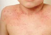
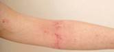
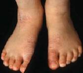
The childhood stage is primarily a papular dermatitis affecting the flexural areas, especially the antecubital and popliteal fossa, wrists, ankles, and neck. Thickened, lichenified plaques with excoriation (Figures 2 to 4) are common. In darker-pigmented children, follicular papules may be the only manifestation. Hypopigmentation and hyperpigmentation can occur, which can cause great anxiety in parents. Pityriasis alba, characterized by hypopigmented, scaly patches on the face, is commonly seen. Keratosis pilaris, or spiny hair follicles, commonly affects the posterior aspects of the upper arms and the anterior thighs.
The adult stage is unpredictable. Affected patients may have had only a few outbreaks since infancy or they may have had a chronic, relapsing course. Hand dermatitis is common and may be the only manifestation of adult atopic dermatitis, which can lead to significant disability. Like affected children, adults also commonly have lichenification of the flexures and facial dermatitis.
The diagnosis of atopic dermatitis depends on a personal and/or family history of atopy coupled with the clinical signs and symptoms described by Hanifin (Table 1).11 Pruritus and xerosis are key elements; without them, the diagnosis should be questioned.9
Major Features (must have 3)
|
Minor or Less-Specific Features
|
Reprinted from Hanifin JM. Atopic dermatitis in infants and children. Pediatr Clin North Am 1991; (4)38:763–789. Copyright © 1991 with permission from Elsevier. https://www.sciencedirect.com/journal/pediatric-clinics-of-north-america
Atopic dermatitis can resemble other types of dermatitis (seborrheic dermatitis, allergic contact dermatitis, irritant contact dermatitis) and dermatophytosis. It may be a component of rare genetic diseases such as Netherton syndrome, ichthyosis, and immunodeficiency syndromes (e.g., X-linked agammaglobulinemia, Wiskott-Aldrich syndrome, isolated IgA deficiency, and severe combined immunodeficiency disease). Helpful diagnostic tests include a serum IgE level, serum protein electrophoresis, fungal scraping for potassium hydroxide preparation and culture, and skin biopsy.
Atopic dermatitis tends to be a chronic relapsing disease. The goals of therapy should be to reduce the number and severity of flares and to increase the number of disease-free periods. The mainstay of treatment for atopic dermatitis is hydrating the skin with the regular use of emollients and suppressing cutaneous inflammation with topical corticosteroids. Topical calcineurin inhibitors have become an important adjunctive therapy. For severe disease, especially during acute flares, systemic corticosteroids may be necessary. Secondary infections require treatment with topical or oral antibiotics, or both. Oral antihistamines can help decrease pruritus. In severe, recalcitrant cases, phototherapy or systemic immunosuppressive medications may be necessary.
Most patients with atopic dermatitis require hydration though the liberal use of bland emollients, which serve to hydrate the stratum corneum and maintain the lipid barrier. Sufficient emollients applied liberally several times a day may be enough to significantly reduce the disease activity of atopic dermatitis. Parents of infants and toddlers should apply a bland emollient to the entire body with each diaper change. Older children should apply bland emollients in the morning, after school, and at bedtime. Bathing should be limited to brief, cool showers once daily. Soap, which dries and irritates the skin, should be avoided, but gentle lipid-free cleansers are beneficial.
Corticosteroids suppress lymphocyte activity in the skin, thereby decreasing inflammation. Patients can use a low-potency topical steroid (hydrocortisone or desonide) for day-to-day control of mild disease and a medium-potency steroid (triamcinolone acetonide, fluticasone, or fluocinolone) for more severe flares. Low-potency topical steroids are suitable for infants and for intertriginous and sensitive areas (face, genitals); more potent steroids should be avoided on these sites. Severe, widespread disease can require systemic corticosteroids. Because of the well-known side effects of systemic corticosteroids (e.g., adrenal suppression, osteoporosis, hypertension, diabetes, obesity, striae), their use should be limited to patients with severe disease.
Topical calcineurin inhibitors (tacrolimus, pimecrolimus) are effective alternatives to the chronic use of topical corticosteroids. Topical calcineurin inhibitors bind calcineurin and block the activation of T cells by cytokines, thus halting the inflammatory cascade that leads to atopic dermatitis. Topical calcineurin inhibitors are especially suitable for more delicate areas such as the face and genitals because they do not carry the risks of atrophy, telangiectasias, and striae associated with the chronic use of steroids. Reports have surfaced suggesting a possible risk of lymphoma associated with high-dose oral pimecrolimus in animal studies,12 prompting the US Food and Drug Administration to put out a black box warning advising against the use of topical calcineurin inhibitors in children younger than 2 years. However, there are no data to support an increased risk of lymphoma with topical treatment in humans.12 Topical calcineurin inhibitors should be used for a limited time and only on affected skin. They should not be used as a daily moisturizer, first-line therapy, or preventive therapy.
The pruritus associated with atopic dermatitis can be severe and often interferes with school, work, and sleep. Despite a lack of objective data to support their use, antihistamines are commonly used to break the itch-scratch-itch cycle.13 Nonsedating antihistamines such as fexofenadine, cetirizine, loratadine, and desloratadine can help offset daytime itching without somnolence. Sedating antihistamines such as diphenhydramine or hydroxyzine are often helpful for nighttime pruritus.
Patients with atopy have an abnormal tolerance to S aureus colonization of the skin, which can exacerbate the dermatitis. Affected patients should use lipid-free antibacterial cleansers. For open wounds, a topical antibiotic such as mupirocin can help to prevent secondary impetiginization (Figures 5 and 6). An oral antibiotic with S aureus coverage and good skin penetration, such as amoxicillin-clavulanic acid, cephalexin, or azithromycin, is necessary for extensive excoriations and impetigo.
In severe, recalcitrant cases, ultraviolet (UV) light treatments (UVB or psoralen plus UVA [PUVA]) and immunosuppressive medications (e.g., methotrexate, cyclosporine, azathioprine, mycophenolate mofetil) may be helpful.13 These should be used very cautiously and with close monitoring and should be reserved for the most severe cases.
Allergic contact dermatitis from topical medications, cosmetics, or metals should be considered in patients with recalcitrant disease. Evaluation by an environmental dermatologist and an allergist, including patch, pinprick, and serum radioallergosorbent testing, may be warranted. Topical medications that are known sensitizers, such as lidocaine, doxepin cream, and diphenhydramine cream, as well as topical antibiotics such as neomycin, should be strictly avoided. Allergic contact dermatitis to topical steroids should be considered in any patient who fails to improve or worsens with the use of topical steroids.
A multitude of new treatments are currently available or under investigation for atopic dermatitis. These range from oral to topical agents and each has an important inflammatory target. Key components of this TH2-mediated disease include cytokines such as IL-4, IL-5, IL-13, and IL-31. Dupilumab binds a subunit of the IL-4 receptor and therefore blocks IL-4 and IL-13. This medication is approved for the treatment of moderate-to-severe atopic dermatitis and there are ongoing long-term studies.14
Similarly, lebrikizumab is an IL-13 antibody in trial that has demonstrated efficacy in atopic patients who were still using topical corticosteroids; continued studies are needed.15
IL-31 seems to play a role in atopic dermatitis and itch development. Nemolizumab (CIM331) targets the IL-31 receptor A and has been shown to decrease itch sensation among affected patients in early reports.16
A related approach includes targeting Janus kinase (JAK) signaling, which plays an important role in immune function and is involved in inflammatory skin conditions including chronic itch. While further investigation remains necessary, early data show JAK inhibitors (jakinibs) to be a promising therapy option.17 JAK assists in the function and effect of various TH2 cytokines discussed above. Baricitinib, an inhibitor of JAK1 and JAK2, has been shown to reduce atopic dermatitis and itch in patients who continued to use topical corticosteroids.18 Additional JAK1 inhibitors currently under study include PF-04965842 and ABT-494 (upadacitinib).17
While the JAK pathway was first targeted with oral tofacitinib citrate (a JAK1 and JAK3 inhibitor)19, topical preparations are in trial including topical tofacitinib.20 In addition, the efficacy and safety of a topical JAK inhibitor called JTE-052—which notably inhibits JAK1, JAK2, JAK3, and tyrosine kinase 2—has been reported with promising results.21 Topical ruxolitinib studies are underway as well.17
Lastly, crisaborole, a topical phosphodiesterase 4 inhibitor, is approved for patients with mild-to-moderate atopic dermatitis and has demonstrated efficacy.22 Targeting this pro-inflammatory molecule has led to the development of other phosphodiesterase 4 inhibitors.23
With novel therapies emerging, it is necessary to continue investigation into the efficacy and safety of these various systemic and topical treatments. Furthermore, studies are overall lacking in the pediatric population. Nonetheless, the future for the treatment of atopic dermatitis is encouraging and rapidly expanding.
Atopic dermatitis is a chronic disease with intermittent flares and spontaneous remissions. Approximately 40% to 60% of children with atopic dermatitis have the disease in adulthood, usually manifested as hand dermatitis. More than 75% of children with atopic dermatitis also have asthma or allergic rhinitis.
With good skin care, including the application of appropriate moisturizers, and the use of topical corticosteroids or topical calcineurin inhibitors, most patients with atopic dermatitis do well.